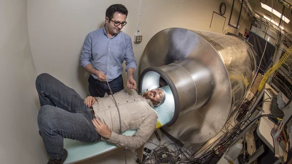
NIH awards $6M grant for noninvasive brain scan helmet
Researchers from Albuquerque, New Mexico-based Sandia National Laboratories have received a new grant to develop a wearable, noninvasive brain scan helmet, the organization announced Thursday.
The federally funded research and development center will use its $6 million National Institutes of Health award to create a more comfortable and, perhaps, more accurate magnetoencephalography imaging prototype.
“This is the future of MEG,” Amir Borna, a lead investigator on the project, said in the announcement.
Typically, clinicians use magnetoencephalography to pinpoint the sources of epilepsy; and researchers often use it to study brain development—notably, for Alzheimer’s disease and stroke insights. In order to achieve this, however, patients must remain still for long periods of time underneath a “helmet-like dome.”
Sandia’s prototype would allow individuals to wear the device, offering more freedom and a more controlled research environment—a particularly promising development for children and people with motor disorders or chronic pain.
“The goal is to expand the number of clinical indications for which MEG may inform clinical care,” said Julia Stephen, an advisor on the project and director of Mind Research Network’s MEG core lab in Albuquerque.
What really separates this project from traditional systems is the team’s use of a new quantum sensor known as an optically pumped magnetometer. These novel devices do not require liquid helium, nor the “rigid” design of commercial systems, but they are just as accurate.
“We have demonstrated a functional brain-imaging system using our quantum sensors that is as reliable as a commercial superconductor-based system,” Borna said.
In the future, the group is planning to scale the number of sensors on its helmet from six, up to 20. Additionally, they want to develop a magnetically shielded room instead of a tube so the subject can move and walk around.
Further details on this project are published in PLOS One.

