Artificial Intelligence

Artificial intelligence (AI) is becoming a crucial component of healthcare to help augment physicians and make them more efficient. In medical imaging, it is helping radiologists more efficiently manage PACS worklists, enable structured reporting, auto detect injuries and diseases, and to pull in relevant prior exams and patient data. In cardiology, AI is helping automate tasks and measurements on imaging and in reporting systems, guides novice echo users to improve imaging and accuracy, and can risk stratify patients. AI includes deep learning algorithms, machine learning, computer-aided detection (CAD) systems, and convolutional neural networks. 
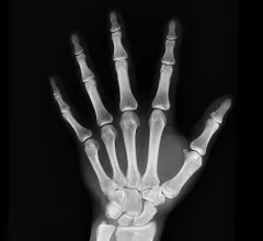
hand x-ray radiograph

A ‘powerful’ tool: Commercially available AI significantly outperforms radiologist reads for trauma imaging

The software was put to the test on nearly 5,000 trauma radiographs, assessing for the presence of fractures, dislocations, elbow effusions and focal bone lesions. In some cases, it outperformed radiologists by as many as 82 points. 

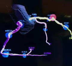
AI-generated coronary tree from a patient's CT scan showing a color code of areas of interest for plaque burden from the Cleerly software shown at SCCT 2022.

VIDEO: Why plaque composition CT imaging may be the future of cardiology

Former European Society of Cardiology president Prof. Jeroen Bax explains the difficulty in quantifying these plaques manually and how artificial intelligence may change this to allow rapid, detailed quantitative analysis. 

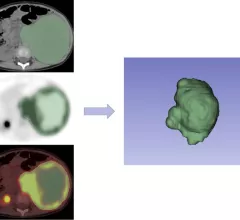
neuroblastoma prognosis

18F-FDG PET/CT radiomics nomogram provides detailed insight into neuroblastoma

Better risk stratification can improve clinical decision making and better outcomes, experts involved in the study explained in EJR.

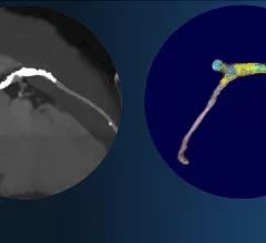
Left, coronary CT angiography of a vessel showing plaque heavy calcium burden. Right, image showing color code of various types of plaque morphology showing the complexity of these lesions. The right image was processed using the FDA cleared, AI-enabled plaque assessment from Elucid.

Cardiac CT soft plaque assessment may offer paradigm shift for coronary disease screening

New artificial intelligence software that can evaluate coronary CT scans to automatically assess soft plaques were by far the biggest technology advance discussed at the Society of Cardiovascular Computed Tomography (SCCT) 2022 meeting. 

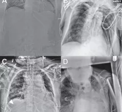
enteric tube placement on radiographs

Algorithm spots enteric tube misplacement on x-ray with great accuracy

The model was externally validated using more than 1,500 radiographs with real-world incidence of critically misplaced tubes. 

Thumbnail

Content-based AI system decreases read times by 31% while increasing accuracy

Using CBIRS decreased interpretation times despite the radiologists who utilized them having to review the additional information the system provided.

Thumbnail

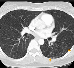
Commercial PE-detecting algorithm identifies incidental clots on CT

Experts involved in the study, which analyzed more than 3,000 CT scans, suggested that there could be a future role for the algorithm to assist radiologists in busy settings.

Aidoc announces strategic hospital partnership that will advance AI in clinical settings

The software will be used for the triage and notification of intracranial hemorrhage (ICH), pulmonary embolism (PE) and c-spine fractures identified on CT imaging.

Around the web

Positron, a New York-based nuclear imaging company, will now provide Upbeat Cardiology Solutions with advanced PET/CT systems and services. 

The nuclear imaging isotope shortage of molybdenum-99 may be over now that the sidelined reactor is restarting. ASNC's president says PET and new SPECT technologies helped cardiac imaging labs better weather the storm.

CMS has more than doubled the CCTA payment rate from $175 to $357.13. The move, expected to have a significant impact on the utilization of cardiac CT, received immediate praise from imaging specialists.