Computed Tomography

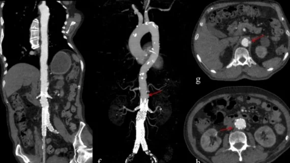
Computed tomography (CT) is a fast and accurate imaging modality often used in emergency settings and trauma imaging. CT scans, with or without (or both) iodinated contrast are frequently used to image the brain, chest, abdomen and pelvis, but also have post-imaging reconstructive capabilities for detailed orthopedic imaging. It is now a standard imaging modality in emergency rooms to quickly assess patients. CT uses a series of X-ray images shot as the gantry rotates around the patient. Computer technology assembles these into into a dataset volume than can be slices on any access, or advanced visualization software can extract specific parts of the anatomy for study. Find more content specific to cardiac CT.
Thumbnail

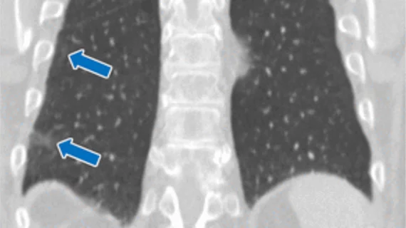
Machine learning uses CT images to predict survival odds of melanoma patients

A unique radiomic signature forecasted overall survival in patients with advanced melanoma and could be used to help providers choose specific therapies.

Thumbnail

Simple quality improvement measures reduce problematic CT overuse for liver imaging

Updated department protocols dropped the median monthly number of abdominopelvic CTs with and without contrast from 74 scans down to 53, experts explained in Current Problems in Diagnostic Radiology.

liver cancer

Deep learning decreases CT radiation dose by 65% in patients with liver metastases

Scans using a smaller dose were accurate when detecting lesions 0.5 cm or larger but more research into low-contrast lesions is still necessary, experts cautioned.

Thumbnail

Bolus tracking with individualized delays for abdominal multiphase CT beats fixed delay protocols

Current bolus tracking technology remains limited by the fixed delay before the start of a scan, experts explained in the European Journal of Radiology.

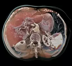
An example of a life-like 3D rendering made from a photon-counting CT scan on the Naeotom Alpha system from Siemens.

First clinical photon-counting CT tops past 50 years of technology, with ‘striking’ improvements

The FDA-cleared machine reduced noise by up to 47% and offers new information that can change patient management, Mayo Clinic experts said.

Thumbnail

‘One-stop shop’ CT protocol boosts definitive diagnostic rates in patients with acute chest pain

The comprehensive approach yields crucial information usually obtained via CT and MRI, which can cut time to diagnosis, experts explained.

Thumbnail

Body CT scans detect infection areas among ICU patients with sepsis

While guidelines stress the importance of quickly eradicating the underlying source of infection, data on the best imaging strategy remains lacking, researchers reported.

RSNA21: Deep learning automatically labels coronary arteries to produce structured CCTA reports

The algorithm yielded a 94.4% accuracy for labeling arteries in accordance with the Society of Cardiovascular Computed Tomography guidelines.

Around the web

Positron, a New York-based nuclear imaging company, will now provide Upbeat Cardiology Solutions with advanced PET/CT systems and services. 

The nuclear imaging isotope shortage of molybdenum-99 may be over now that the sidelined reactor is restarting. ASNC's president says PET and new SPECT technologies helped cardiac imaging labs better weather the storm.

CMS has more than doubled the CCTA payment rate from $175 to $357.13. The move, expected to have a significant impact on the utilization of cardiac CT, received immediate praise from imaging specialists.