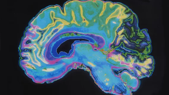
A new MRI technique allows physicians to superimpose images of tiny veins in the brain over conventional images of brain lesions, potentially helping to identify multiple sclerosis (MS) and reduce misdiagnosis, according to a Cedars-Sinai statement.
Misdiagnosis of MS is a common problem, mainly because it is often identified by the appearance of white spots on MRI brain scans. Although white spots can indeed be an indicator of brain lesions commonly caused by MS, they can also result from a number of other common conditions, such as migraines.
“Diagnosis is complex because many other diseases mimic MS, and while we have a set of diagnostic criteria, there’s no single test that is definitive,” said MS specialist Marwa Kaisey, MD, an assistant professor of Neurology at Cedars-Sinai.
MS lesions do have one major distinguishing factor: they often form around tiny veins in the brain known as veinules, which serve as entry points to attack brain tissue. However, it was previously impossible to see whether or not there was a vein in the middle of a lesion on an MRI.
Now, pioneered by Pascal Sati, PhD, director of the Neuroimaging Program in the Department of Neurology and associate professor of Neurology at Cedars-Sinai, the new technique uses MRI sequences which allow physicians to capture images of the brain’s veinules. When superimposed over conventional MRI images of the lesions, Sati said, it becomes easy to determine whether any brain lesions are formed around a central vein—known as “central vein sign”—and therefore are likely caused by MS.
“With the central vein sign, we can clearly see which lesions are related to MS,” Sati said. “This information is empowering doctors to make decisions about whether to continue a patient’s current therapy, switch to a different MS therapy, or treat them for a completely new, or different, condition.”
Sati is also developing a machine-learning algorithm to identify the central vein sign on MRIs and alert physicians to it.
Related Multiple Sclerosis Content:
New MRI technique earlier detects multiple sclerosis, potentially improving treatment approaches
AI pinpoints 3 multiple sclerosis subtypes using thousands of brain MRI scans
7T MRI reveals new view of damage in multiple sclerosis patients
Applying deep learning to PET/CT scans helps clinicians diagnose neurodegenerative disorders