Magnetic Resonance Imaging

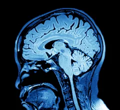
Magnetic resonance imaging (MRI) is the gold standard imaging modality for soft tissues. It produces detail cross-sectional images of soft tissue and bone anatomy, including muscles, tendons, ligaments, brain and organs, without the use of ionizing radiation. In addition to orthopedic imaging, MRI is also used for heart, brain and breast. MRI uses gadolinium contrast in many exams to highlight tissues and blood vessels, which enhances images and offers better diagnostic quality. It can also be used in conjunction with PET scans. How does MRI work? MR creates images by using powerful magnets to polarize hydrogen atoms in water (the body is made of of more than 80% water) so they face in one direction. A radiofrequency pulse is then used to ping these atoms, causing them to wobble, or resonate. The MRI coils detect this and computers can assemble images from the signals. Basic MRI scans will focus on the resonance of fat and water in two different sequences, which highlight and contrast different features in the anatomy.

Thumbnail

Radiologists must embrace low-field MRI or ‘we will be in trouble,’ expert says

The specialty needs to work together and address a number of issues surrounding the disruptive technology, Yale Director of MRI Services Jeffrey C. Weinreb, MD, argued.

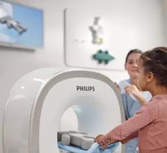
Cooperation during pediatric MRIs improves with parental participation, study shows

Parent-assisted MRI scans improve imaging procedures and reduce associated costs, experts reported recently.

Thumbnail

Providers are requesting fewer preoperative MRIs for breast cancer patients

Female surgeons, in particular, are abiding by evidence-based guidelines, but a more selective approach is still needed among doctors.

heart vaccine myocarditis covid-19

Are COVID mRNA vaccines a cause of myocarditis in adolescents? MRI findings hold clues

Scan data published in the American Journal of Roentgenology suggests radiologists must remain cognizant of the link between myocarditis and vaccines.

liver cancer

MRI biomarkers less invasive, more accurate option for identifying deadly liver disease

The findings present positive news for patients who are looking to avoid the discomfort of a biopsy in the pursuit of a diagnosis, experts explained recently.

Thumbnail

Radiologists see potential to reduce GBCA administration with new synthetic MRI technique

Even though synthetic samples may not be perfect copies of original images, experts say they can act as a useful substitute for gadolinium-enhanced exams.

Thumbnail

Advanced MRI shows potential for assessing asymptomatic patients at risk for Alzheimer’s

Experts compared people with a genetic mutation associated with the disease and non-carriers, sharing their new insights in Radiology.

Health giant launches ‘coaching’ solution to help patients with MRI anxiety

Company officials believe the new offerings can enhance quality and lead to more sedation-free exams. 

Around the web

Positron, a New York-based nuclear imaging company, will now provide Upbeat Cardiology Solutions with advanced PET/CT systems and services. 

The nuclear imaging isotope shortage of molybdenum-99 may be over now that the sidelined reactor is restarting. ASNC's president says PET and new SPECT technologies helped cardiac imaging labs better weather the storm.

CMS has more than doubled the CCTA payment rate from $175 to $357.13. The move, expected to have a significant impact on the utilization of cardiac CT, received immediate praise from imaging specialists.