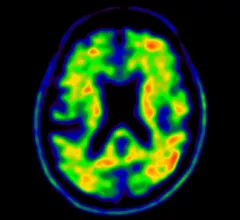
Molecular Imaging

Molecular imaging (also called nuclear medicine or nuclear imaging) can image the function of cells inside the body at the molecular level. This includes the imaging modalities of positron emission computed tomography (PET) and single photon emission computed tomography (SPECT) imaging. How does PET and SPECT imaging work? Small amounts of radioactive material (radiopharmaceuticals) injected into a patient. These can use sugars or chemical traits to bond to specific cells. The radioactive material is taken up by cells that consume the sugars. The radiation emitted from inside the body is detected by photon detectors outside the body. Computers take the data to assemble images of the radiation emissions. Nuclear images may appear fuzzy or ghostly rather than the sharper resolution from MRI and CT.  But, it provides metabolic information at a cellular level, showing if there are defects in the function of the heart, areas of very high metabolic activity associated with cancer cells, or areas of inflammation, data not available from other modalities. These noninvasive imaging exams are used to diagnose cancer, heart disease, Alzheimer’s and Parkinson’s disease, bone disorders and other disorders. 

Researchers create novel method predicting tumor response in radioligand therapy

A team of German researchers used PET/CT and modeling practices to create a new method to pattern and predict tumor response in radioligand therapy (RLT), which authors say is the first attempt to do so.

Thumbnail

Could a breast imaging pill eliminate the need for annual mammograms?

The "disease screening pill," developed by researchers from the University of Michigan, lit up malignant tumors in mice when exposed to near-infrared light—all without the use of radiation, according to a study published in Molecular Pharmaceutics.

Thumbnail

SNMMI collaborates with industry leaders to advance molecular imaging

On May 17, the Society of Nuclear Medicine and Molecular Imaging (SNMMI) announced the Value Initiative Industry Alliance—a collaboration among corporate community members that will help implement the organization’s Value Initiative and advance nuclear medicine.

Thumbnail

PET tracer may predict response to therapy in bone-dominant breast cancer patients

A prospective comparison study of two PET tracers found 18F-FDG could provide valuable measures of activity in bone-dominant (BD) metastatic breast cancer (MBC) patients, while also predicting responses to therapy.

Thumbnail

NIH funds development of 3 national cryo-electron microscopy centers

The National Institutes of Health (NIH) announced efforts to widen the scientific community's access to cryo-electron microscopy (cryo-EM) through its Transformative High Resolution Cryo-Electron Microscopy program.  

Thumbnail

Canadian particle accelerator produces Tc-99m isotopes

A team at the University of Alberta in Canada has devised a method utilizing a cyclotron particle accelerator to produce the radioactive tracer technetium-99m—the parent of Molybdenum-99. It may be able to produce enough radioactive isotope for the entire province, CBC.ca reports.

Thumbnail

NIH merges microscopes to generate clearer images of processes inside cells

Scientists at the National Institute of Biomedical Imaging and Bioengineering (NIBIB) announced they had merged two microscope technologies to generate clearer images of rapid processes occurring inside human cells.

Thumbnail

Tau PET tracer found effective in 1st in-human study of its kind

A team of Johns Hopkins University researchers—conducting the first in-human PET study of three novel tau radiopharmaceuticals in Alzheimer’s disease patients—found [18F]RO-948 was the most capable for characterizing tau pathology in the disease.

Around the web

Positron, a New York-based nuclear imaging company, will now provide Upbeat Cardiology Solutions with advanced PET/CT systems and services. 

The nuclear imaging isotope shortage of molybdenum-99 may be over now that the sidelined reactor is restarting. ASNC's president says PET and new SPECT technologies helped cardiac imaging labs better weather the storm.

CMS has more than doubled the CCTA payment rate from $175 to $357.13. The move, expected to have a significant impact on the utilization of cardiac CT, received immediate praise from imaging specialists.