Molecular Imaging

Molecular imaging (also called nuclear medicine or nuclear imaging) can image the function of cells inside the body at the molecular level. This includes the imaging modalities of positron emission computed tomography (PET) and single photon emission computed tomography (SPECT) imaging. How does PET and SPECT imaging work? Small amounts of radioactive material (radiopharmaceuticals) injected into a patient. These can use sugars or chemical traits to bond to specific cells. The radioactive material is taken up by cells that consume the sugars. The radiation emitted from inside the body is detected by photon detectors outside the body. Computers take the data to assemble images of the radiation emissions. Nuclear images may appear fuzzy or ghostly rather than the sharper resolution from MRI and CT.  But, it provides metabolic information at a cellular level, showing if there are defects in the function of the heart, areas of very high metabolic activity associated with cancer cells, or areas of inflammation, data not available from other modalities. These noninvasive imaging exams are used to diagnose cancer, heart disease, Alzheimer’s and Parkinson’s disease, bone disorders and other disorders. 
Thumbnail

SNMMI urges Aetna to alter coverage of 2 PET tracers

The Society of Nuclear Medicine and Molecular Imaging (SNMMI) addressed two letters to Aetna recommending the health insurance giant alter its radiopharmaceutical coverage policy to include PET imaging agents dotatate and fluciclovine.

Thumbnail

Homemade microscope shows cancer virus clinging to human DNA

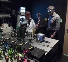
A high-tech microscope developed by scientists at the University of Virginia (UVA) School of Medicine has captured images of cancer-causing viruses clinging to human DNA, according to a UVA Health System release.

Thumbnail

MRI shows immune cells healing brain's lining after a concussion

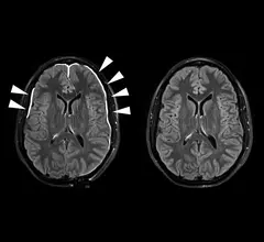
According to a National Institutes of Health (NIH) news release published April 17, NIH scientists observed in real time how immune system cells cooperatively fix the damaged lining of the brain after a concussion.

Study finds brain continues to produce new cells into old age, contradicting recent findings

Columbia University researchers recently found that the human brain continues to produce hundreds of new neurons every day, even into old age, according to an article by the Los Angeles Times.

The case for modernizing biodistribution reporting in nuclear medicine

Radiopharmaceutical dosimetry (RD) is an integral part of nuclear imaging and therapy, but current standards for documenting and reporting compound travel patterns inside the human body—biodistribution—in dosimetry-related studies do not exist.

Monitoring cerebrospinal fluid biomarkers, imaging of brain injuries may identify abnormal neurodevelopment

Monitoring cerebrospinal fluid (CSF) biomarkers and imaging for genetic mediators of brain injuries in children may help identify those at risk for abnormal brain development, according to a study published in the Journal of the American Medical Association.

SPECT/CT may save more than $1.7M over 3 years in total knee replacement surgeries

By 2030, some 3.5 million Americans are expected to be living with total knee replacements. The price of performing total-knee arthroplasty (TKA) revision surgery continues to rise, costing 60 percent more than the initial replacement. Researchers set out to identify a more cost-effective method.

Thumbnail

Diamonds are an MRIs best friend, may increase imaging sensitivity

A new molecular technique using modified diamonds could increase the sensitivity of an MRI and improve patient diagnosis, according to a University of Melbourne press release.

Around the web

Positron, a New York-based nuclear imaging company, will now provide Upbeat Cardiology Solutions with advanced PET/CT systems and services. 

The nuclear imaging isotope shortage of molybdenum-99 may be over now that the sidelined reactor is restarting. ASNC's president says PET and new SPECT technologies helped cardiac imaging labs better weather the storm.

CMS has more than doubled the CCTA payment rate from $175 to $357.13. The move, expected to have a significant impact on the utilization of cardiac CT, received immediate praise from imaging specialists.