Molecular Imaging

Molecular imaging (also called nuclear medicine or nuclear imaging) can image the function of cells inside the body at the molecular level. This includes the imaging modalities of positron emission computed tomography (PET) and single photon emission computed tomography (SPECT) imaging. How does PET and SPECT imaging work? Small amounts of radioactive material (radiopharmaceuticals) injected into a patient. These can use sugars or chemical traits to bond to specific cells. The radioactive material is taken up by cells that consume the sugars. The radiation emitted from inside the body is detected by photon detectors outside the body. Computers take the data to assemble images of the radiation emissions. Nuclear images may appear fuzzy or ghostly rather than the sharper resolution from MRI and CT.  But, it provides metabolic information at a cellular level, showing if there are defects in the function of the heart, areas of very high metabolic activity associated with cancer cells, or areas of inflammation, data not available from other modalities. These noninvasive imaging exams are used to diagnose cancer, heart disease, Alzheimer’s and Parkinson’s disease, bone disorders and other disorders. 

Thumbnail

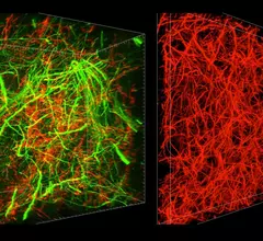
New technique allows for zooming in, out of brain imaging at different levels

A new study published in the journal Nature Biotechnology outlines a new method for brain imaging from researchers at the Massachusetts Institute of Technology (MIT). 

Mouse study shows tumors tamed by T cells targeting two different antigens

TanCAR T cells—so called because they can bind, in tandem, the chimeric antigen receptor (CAR) and both the antigens it targets, HER2 and IL13Ra2—have shown intriguing potential to give oncologists better control over brain tumors.

Calcium molecule linked to aggressive cancer, imaging technique shows

New research shows a particular calcium molecule is linked to aggressive cancers when it stops regulating itself properly, and researchers at Columbia University Medical Center are looking for ways to fix it, according to a recent report.

HTG Molecular Diagnostics Announces VERI/O Laboratory Service

TUCSON, Ariz., June 23, 2016 (GLOBE NEWSWIRE) -- HTG Molecular Diagnostics, Inc. (Nasdaq:HTGM), a provider of instruments and reagents for molecular profiling applications, today announced the introduction of its Tucson‑based VERI/O laboratory service, offering laboratory support for its biopharma clients in biomarker research and companion diagnostic development.  The VERI/O laboratory service formalizes and expands HTG’s traditional service offerings, including molecular profiling of retrospective cohorts to support development of targeted and immuno-oncology therapies, building custom research-use-only (RUO) assays to support early stage clinical programs, and developing companion diagnostic assays for use in Phase III registration trials.

MR Solutions offers choice of two preclinical cryogen-free MRI scanner ranges

MR Solutions, which introduced cryogen-free MRI scanners in 2012, is now offering a choice of two cryogen-free preclinical scanner ranges – the Flexiscan and Powerscan. The Flexiscan range is available with multi-modality accessories which can be mixed and matched according to research requirements. It is a flexible MRI system as it can incorporate integrated multi-modality options such as PET or SPECT. The Flexiscan system requires no specialist knowledge and can be operated simply by running pre-defined settings. There is no need to materially alter the parameters of the machine.

Device with sci-fi name may make molecular-imaging systems better and safer

With a name like “variable angle slant hole collimator,” it’d better be impressive. 

Non-imaging MR application helps head off sepsis in children

An MR-based system for identifying pediatric cases of Candida infection has been shown capable of using low-volume blood specimens to efficiently diagnose or rule out candidemia, the deadliest form of sepsis-causing bloodstream infection.

Nanomed advance may fight brain tumors long considered death sentences

Researchers at the Medical University of South Carolina’s Center for Biomedical Imaging have engineered a lipid nanocarrier capable of carrying a concentrated dose of the chemotherapy drug TMZ directly into the cells of aggressive glioblastoma brain tumors. 

Around the web

Positron, a New York-based nuclear imaging company, will now provide Upbeat Cardiology Solutions with advanced PET/CT systems and services. 

The nuclear imaging isotope shortage of molybdenum-99 may be over now that the sidelined reactor is restarting. ASNC's president says PET and new SPECT technologies helped cardiac imaging labs better weather the storm.

CMS has more than doubled the CCTA payment rate from $175 to $357.13. The move, expected to have a significant impact on the utilization of cardiac CT, received immediate praise from imaging specialists.