Molecular Imaging

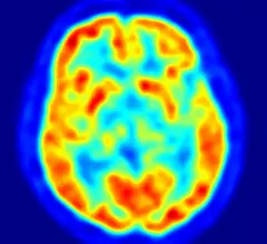
Molecular imaging (also called nuclear medicine or nuclear imaging) can image the function of cells inside the body at the molecular level. This includes the imaging modalities of positron emission computed tomography (PET) and single photon emission computed tomography (SPECT) imaging. How does PET and SPECT imaging work? Small amounts of radioactive material (radiopharmaceuticals) injected into a patient. These can use sugars or chemical traits to bond to specific cells. The radioactive material is taken up by cells that consume the sugars. The radiation emitted from inside the body is detected by photon detectors outside the body. Computers take the data to assemble images of the radiation emissions. Nuclear images may appear fuzzy or ghostly rather than the sharper resolution from MRI and CT.  But, it provides metabolic information at a cellular level, showing if there are defects in the function of the heart, areas of very high metabolic activity associated with cancer cells, or areas of inflammation, data not available from other modalities. These noninvasive imaging exams are used to diagnose cancer, heart disease, Alzheimer’s and Parkinson’s disease, bone disorders and other disorders. 
Thumbnail

Specialists, PET centers now able to register for IDEAS study

The Imaging Dementia – Evidence for Amyloid Scanning (IDEAS) study is now accepting registrations from dementia specialists and PET imaging centers looking to participate in the research.

Thumbnail

Mobile molecular breast imaging rolls into Wisconsin

Marshfield Clinic Health System in Wisconsin has acquired technology that, it says, will make it the world’s first provider of mobile molecular breast imaging (MBI) services.

Thumbnail

New metabolic PET tracer finds deep brain tumors, helps monitor treatment

Stanford University researchers have developed a molecular tracer capable of locating and subsequently following a cancer-elevated protein in hard-to-find brain tumors. 

Pumpkin-shaped molecule said to provide ‘stunning’ MRI contrast

European researchers have discovered a class of molecule that, when exposed to the inert gas xenon, enables MRI contrast significantly sharper than previous contrast techniques.

Preop PET/CT staging for melanoma has high false-positive rate

The use of preoperative PET/CT staging to detect clinically occult metastatic melanoma in patients with sentinel lymph node-positive melanoma has a high false-positive rate and a minimal effect on management, according to a research letter published in JAMA Oncology.

WMIS Presents First Annual Commercial Innovation of the Year Award to MILabs for Breakthrough in SPECT Imaging at WMIC 2015

The World Molecular Imaging Society (WMIS) presented the first annual Commercial Innovation of the Year Award at the 2015 World Molecular Imaging Congress (WMIC) to Frederik Beekman, CEO/CSO, MILabs for his work developing G-SPECT.   

Thumbnail

New whole-body PET probe ferrets out hidden blood clots

A preclinical trial of a novel radiopharmaceutical has shown a single intravenous injection of the probe, when used with whole-body PET, is capable of flagging blood clots wherever in the body they travel and hide. 

NorthStar, Westinghouse partner to produce medical radioisotopes

NorthStar Medical Radioisotopes and Westinghouse Electric have completed a memorandum of understanding under which the companies will explore the potential for producing medical radioisotopes in commercial nuclear reactors.  

Around the web

Positron, a New York-based nuclear imaging company, will now provide Upbeat Cardiology Solutions with advanced PET/CT systems and services. 

The nuclear imaging isotope shortage of molybdenum-99 may be over now that the sidelined reactor is restarting. ASNC's president says PET and new SPECT technologies helped cardiac imaging labs better weather the storm.

CMS has more than doubled the CCTA payment rate from $175 to $357.13. The move, expected to have a significant impact on the utilization of cardiac CT, received immediate praise from imaging specialists.