X-ray

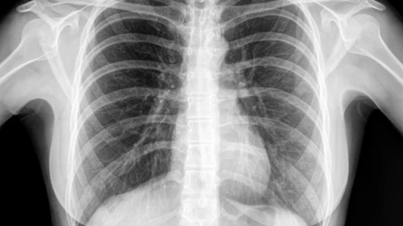
Medical X-rays have mostly converted from film, to computed radiography (CR) that used individual plates to record each X-ray digitally that then had to be uploaded into a PACS, to what is the standard-of-care today with digital radiography (DR). DR X-ray which allows immediate digital transfer of images into a PACS for immediate review. X-rays are used to diagnose fractures, bone abnormalities, lung pathologies and tumors, as well as monitor pediatric growth, plan for surgery and treat oncology patients during radiation therapy. More detailed anatomical imaging, especially soft tissue imaging, is usually sent for advanced imaging with CT or MRI. X-ray, especially mobile DR systems, are a primary use case for artificial intelligence (AI) integration. 
Carestream Horizon X-ray system

Carestream unveils compact X-ray for facilities on a budget

The system is designed for small healthcare facilities, fitting into most rooms without requiring additional ceiling support.

mermaid

Radiology students to run scans on 'mermaid'

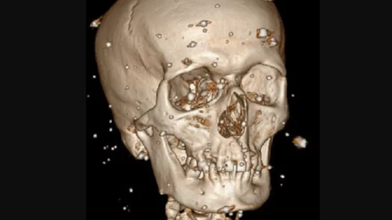
A group of radiology students at Northern Kentucky University are conducting an analysis of the remains of a mysterious artifact known as a "Fiji Mermaid."

artificial intelligence robot evaluates healthcare data

'Transformer' AI model can read chest X-rays like a human radiologist

Researchers in Germany created an AI model that outperforms others in diagnostic accuracy when given access to clinical data that goes beyond an X-ray image.

radiology trends lungs imaging graphs

Radiologists deliver fewer false-positive results than advanced AI models

A new study out of Denmark reveals humans have a definite advantage when a diagnosis requires real-world experience. 

X-ray results suggest telehealth may be a bad fit for treating musculoskeletal conditions

Musculoskeletal X-rays ordered during telemedicine appointments had a higher likelihood of showing "normal" results than those ordered during in-person visits, suggesting those exams may have not been necessary in the first place. 

lung cancer pulmonary nodule chest

FDA clears AI-powered software that pinpoints suspicious findings on chest X-rays

VisiRad XR was developed using curated data sets from across the globe, developer Imidex said in an announcement. 

Artificial intelligence (AI) model using chest x-rays to evaluate cardiac function

No echocardiography, no problem? AI evaluates cardiac function using chest X-rays

Researchers think their new AI model could be especially helpful in areas where qualified physicians and/or ultrasound technology are in short supply.

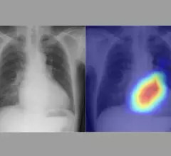
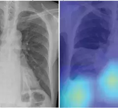
A figure from the study shows a chest radiograph with an area of consolidation involving right lower lung consistent with pneumonia, as well as right pleural effusion. The deep-learning model predicted risk of 30-day mortality of 9%. Right: Gradient- weighted class activation map shows that model prediction was influenced by separate area of image corresponding with heart and liver (yellow and light blue colors). Patient’s CURB-65 score was 4. Patient recovered from pneumonia and remained alive. AJR Image

Deep learning predicts pneumonia mortality on chest X-rays

AI was able to predict 30-day mortality risk predictions more accurately that the current risk assessment.

Around the web

GE HealthCare's flurpiridaz, the PET radiotracer that recently received FDA approval, offers several key benefits over SPECT. Jamshid Maddahi, MD, discussed the details in an exclusive interview. 

Ultrafast MCE could go on to become a go-to treatment option for obstructive coronary artery disease, according to the authors of a new first-in-human clinical study.

Elucid's PlaqueIQ was trained to turn CCTA images into interactive 3D reports that help physicians visualize the presence of atherosclerosis.

Trimed Popup
Trimed Popup