Magnetic Resonance Imaging

Magnetic resonance imaging (MRI) is the gold standard imaging modality for soft tissues. It produces detail cross-sectional images of soft tissue and bone anatomy, including muscles, tendons, ligaments, brain and organs, without the use of ionizing radiation. In addition to orthopedic imaging, MRI is also used for heart, brain and breast. MRI uses gadolinium contrast in many exams to highlight tissues and blood vessels, which enhances images and offers better diagnostic quality. It can also be used in conjunction with PET scans. How does MRI work? MR creates images by using powerful magnets to polarize hydrogen atoms in water (the body is made of of more than 80% water) so they face in one direction. A radiofrequency pulse is then used to ping these atoms, causing them to wobble, or resonate. The MRI coils detect this and computers can assemble images from the signals. Basic MRI scans will focus on the resonance of fat and water in two different sequences, which highlight and contrast different features in the anatomy.
MRA for pulmonary embolus

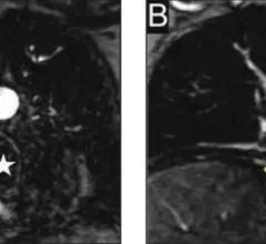
MR angiography a suitable alternative to CT when ruling out pulmonary embolus

The modality switch became especially important during the iodinated contrast shortage of 2022 when clinics were forced to deploy mitigation tactics as a means of preserving their contrast supply. 

June 22, 2023
Covid Mask

New MRI technique offers detailed view of COVID's impact on the brain

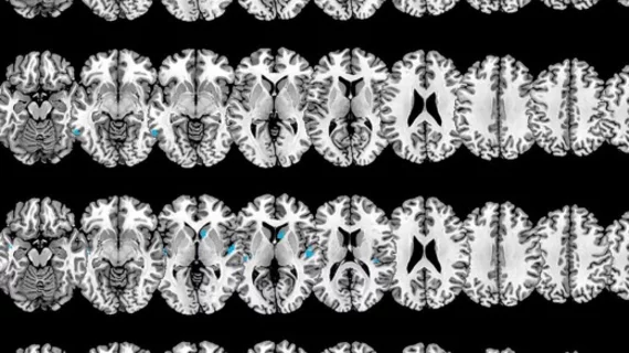
The new technique mixes MRI signals at different gradient pulse strengths and timings, offering better visualization of differences in the way water molecules move through tissues.  

June 14, 2023
gadolinium, GBCAs

When GBCA use in musculoskeletal imaging is and is not appropriate

Although gadolinium-based contrast agents are largely considered safe and are routinely used for MRI exams, experts suggest that providers should still utilize GBCAs sparingly for musculoskeletal studies.

June 14, 2023
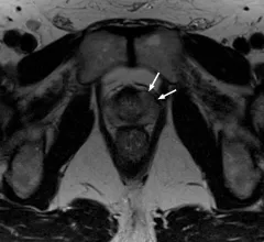
An international panel of experts recently developed and validated a reporting assessment scoring system that analyzes the location and extent of prostate cancer recurrence. 

Prostate cancer size is frequently underestimated on imaging

Newly published data recently revealed just how common the extent of prostate cancer is underestimated based on imaging measurements. 

June 12, 2023
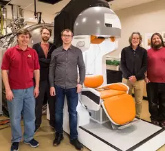
University of Minnesota team develops compact portable MRI. Michael Garwood, second from right, and collaborators at the University of Minnesota created a mobile MRI prototype that could provide diagnostic imaging to rural and underserved populations. Image courtesy of the University of Minnesota.

Curbside MRI: Experts are developing system small enough to fit in a pickup

Someday, getting an MRI exam could be as simple as having food delivered to your door—at least that is the hope of a group of experts at the University of Minnesota who are working on a compact system said to be small enough to sit in the bed of a truck.

June 7, 2023
Philips Healthcare booth sign at HIMSS23.

Philips' latest collaboration could make xenon MR imaging more widely available

In alliance with Polarean, Philips is rolling out a new MR system with advanced multi-nuclei imaging capabilities that will offer radiologists a detailed evaluation of lung ventilation using xenon gas as a contrast agent.

June 7, 2023
stroke

Incomplete neurovascular imaging work-ups to blame for subsequent stroke in patients with TIA

Up to 37% of patients presenting with transient ischemic attack do not receive a full neurovascular imaging work-up, according to new data published in the American Journal of Roentgenology. 

June 1, 2023

AI tool helps identify 'invisible' head injuries on MRIs of college athletes

The tool uses a machine learning technique to identify changes on brain MRIs that would otherwise be overlooked by radiologists due to the subtlety of alterations. 

June 1, 2023

Around the web

Automated AI-generated measurements combined with annotated CT images can improve treatment planning and help referring physicians and patients better understand their disease, explained Sarah Jane Rinehart, MD, director of cardiac imaging with Charleston Area Medical Center.

Two advanced algorithms—one for CAC scores and another for segmenting cardiac chamber volumes—outperformed radiologists when assessing low-dose chest CT scans. 

"Gen AI can help tackle repetitive tasks and provide insights into massive datasets, saving valuable time," Thomas Kurian, CEO of Google Cloud, said Tuesday. 

Trimed Popup
Trimed Popup