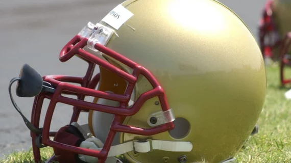
AI tool helps identify 'invisible' head injuries on MRIs of college athletes
A new artificial intelligence tool is helping experts to better understand the implications of “invisible” brain damage in athletes who sustain repetitive blows to the head.
The tool uses a machine learning technique to identify changes on brain MRIs that would otherwise be overlooked by radiologists due to the subtlety of alterations. Using the tool, a team of researchers in the Department of Radiology at NYU Grossman School of Medicine were able to distinguish between the brains of athletes who play contact sports and those who participate in noncontact sports based on their imaging alone.
Experts detailed their findings recently in The Neuroradiology Journal.
“Our findings uncover meaningful differences between the brains of athletes who play contact sports compared to those who compete in noncontact sports,” the study’s senior author Yvonne Lui, MD, who is a neuroradiologist at New York University Grossman School of Medicine, and co-authors noted. “Since we expect these groups to have similar brain structure, these results suggest that there may be a risk in choosing one sport over another.
For their work, the team analyzed whole-brain MRIs from 36 college athletes who played contact sports (mostly football) and 45 noncontact athletes. All were male, and none had sustained concussions prior to undergoing imaging.
The tool was trained to identify certain features in brain tissue that would indicate that a head injury had occurred. Through this, the team was able to identify two MRI metrics that most accurately discriminate between the two groups of athletes—mean diffusivity and mean kurtosis.
Mean diffusivity analyzes the flow of water through brain tissues, while mean kurtosis assesses the microstructure, particularly in areas known to be involved with emotions, memory and learning. The AI tool identified alterations in both of these metrics in contact sport athletes.
“Our results highlight the power of artificial intelligence to help us see things that we could not see before, particularly ‘invisible injuries’ that do not show up on conventional MRI scans,” the group explained. “This method may provide an important diagnostic tool not only for concussion, but also for detecting the damage that stems from subtler and more frequent head impacts.”
The team has plans to use the same technique to study head injuries in female athletes in the future.
The study abstract is available here.

