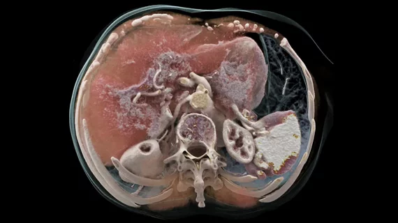
First clinical photon-counting CT tops past 50 years of technology, with ‘striking’ improvements
The first photon-counting CT system made available for clinical use offers several advantages over top-of-the-line CT modalities, including higher image quality and less radiation exposure.
In late September, the U.S. Food and Drug Administration cleared Siemens’ Naeotom Alpha photon-counting computed tomography (CT) system, calling it the first major upgrade to CT technology in nearly a decade. Mayo Clinic researchers recently evaluated the system in a handful of patients and their results support the FDA’s high praise.
Prospective findings showed the machine achieved better spatial resolution compared to current systems along with improved noise properties. Photon-counting reduced noise by up to 47% and dropped radiation dose by 30%, experts reported Tuesday in Radiology.
“Photon-counting-detector CT is so exciting because it provides information that existing CT detectors—the types that we’ve used for over 50 years—previously just couldn’t capture,” Cynthia McCollough, PhD, director of Mayo’s CT Clinical Innovation Center, added. “For many clinical applications, the improvements in image quality are really striking.”
For their investigation, McCollough teamed up with employees at Siemens Healthineers to test the technical capabilities of the new CT machine in four participants. They chose to perform CT angiography of the coronary arteries and abdomen/pelvis, whole-body low-dose CT for skeletal surveillance and temporal bone imaging between May and August of this year.
Using the novel device in high-resolution mode resulted in a 125-micron in-plan spatial resolution and 0.3 mm longitudinal resolution, the smallest ever recorded for a clinical CT modality, the authors noted.
Overall, photon-counting-detector CT outperformed even the top computed tomography devices, the experts added. In those with coronary artery disease, for example, it offers an image quality that is difficult to accomplish via traditional CT scanning.
“Every time that CT technology has advanced to provide new technical capabilities, new clinical applications have followed,” McCollough explained. “With PCD-CT, we are seeing anatomy and disease at a resolution that has never before been achieved in patients, and that is providing physicians new information and insights—insights that can change patient management.”
VIDEO: Example of photo-counting cardiac CT with calcified coronaries

