Positron emission tomography/computed tomography is a hybrid nuclear medicine imaging technique that helps radiologists spot abnormal metabolic activity. PET/CT is commonly used to diagnose cancers, heart diseases and certain brain disorders, among other conditions.
Experts hope the information gained from their research could help providers better determine whether patients will regain mobility after sustaining an injury.
If 25% tariffs go into effect, it could have a big impact on the cost of medical imaging and radiotherapy systems, with many manufacturing facilities in Mexico.
There are numerous radiotracers specifically designed to identify prostate cancer, but experts say one may be superior to the others at both the patient and lesion level.
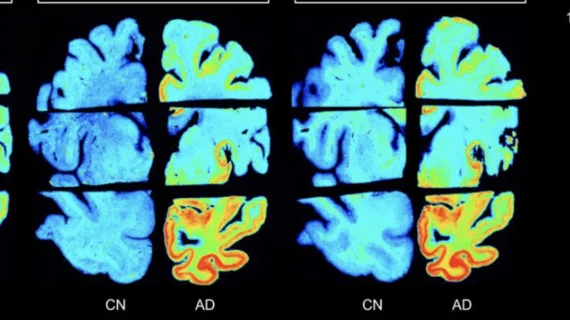
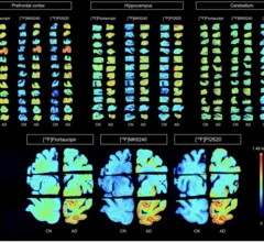
The new appropriate use criteria define 17 specific clinical scenarios, guiding providers on situations when amyloid or tau imaging are and are not appropriate.