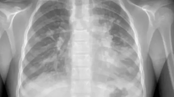
X-ray

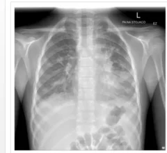
Medical X-rays have mostly converted from film, to computed radiography (CR) that used individual plates to record each X-ray digitally that then had to be uploaded into a PACS, to what is the standard-of-care today with digital radiography (DR). DR X-ray which allows immediate digital transfer of images into a PACS for immediate review. X-rays are used to diagnose fractures, bone abnormalities, lung pathologies and tumors, as well as monitor pediatric growth, plan for surgery and treat oncology patients during radiation therapy. More detailed anatomical imaging, especially soft tissue imaging, is usually sent for advanced imaging with CT or MRI. X-ray, especially mobile DR systems, are a primary use case for artificial intelligence (AI) integration. 
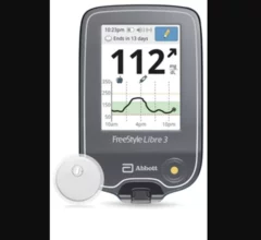
Abbott’s FreeStyle Libre 3 integrated continuous glucose monitoring (iCGM) system

Abbott CGM sensors can now be worn during common imaging procedures

Sensors from the FreeStyle Libre 2 and Libre 3 continuous glucose monitoring systems can now be worn during X-rays, CT scans and MRI scans. The news represents a shift in policy from the FDA, one that came after the agency reviewed extensive testing data.

artificial intelligence healthcare industry digest

ChatGPT is overly worried about ED patients

The popular AI chatbot tends to over-order X-rays, prescribe too many antibiotics and admit too many patients to the hospital when compared with a resident emergency department physician. 

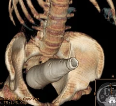
CT scan of a Coca-Cola bottle rectal foreign body. There are a sizable number of emergency room cases each year for items people insert into their rectums that become lodged or lost inside the colon. This includes many types of household items beyond vibrators and commercially made butt plugs. Ghonaim E, Rectal foreign body. Case study, Radiopaedia.org.

PHOTO GALLERY: Abdominal, pelvic and rectal foreign bodies

This is a clinical photo gallery showcasing the uncommon findings of foreign bodies in pelvic and abdominal CT and X-ray, often from emergency room visits. 

virtual reality surgical planning

Rad techs helpfully ‘stand in a surgeon’s shoes’ thanks to virtual reality

Johns Hopkins researchers demonstrated the worthiness of the concept this year.

Thumbnail

Northwestern Medicine is collaborating with Dell to develop AI for reading X-rays

The new artificial intelligence model is currently reading draft radiographs at the Illinois health system.

john_constable

Scientists turn to medical imaging for deep analysis of John Constable painting

Researchers from the University of Bradford deployed CT, Raman spectroscopy, 3D microscopy and an X-ray to confirm the authenticity of one of Constable's landscapes.

Thumbnail

SIR 2024: UAE can eliminate the need for hysterectomy after postpartum hemorrhage

In a study presented at the Society of Interventional Radiology Annual Scientific Meeting, the procedure reduced hysterectomies by 94%.

Head CT of a soldier with shrapnel wounds to head at a hospital in Dnipro. Photo by radiologist Olga Kachanova. Courtesy of RSNA.

PHOTO GALLERY: Radiology images of Ukrainian trauma patients wounded in war

A collection of medical images from hospitals in Ukraine showing radiology equipment in use and clinical images of wounded soldiers and civilians.


Around the web

The two companies aim to improve patient access to high-quality MRI scans by combining their artificial intelligence capabilities.

Positron, a New York-based nuclear imaging company, will now provide Upbeat Cardiology Solutions with advanced PET/CT systems and services. 

The nuclear imaging isotope shortage of molybdenum-99 may be over now that the sidelined reactor is restarting. ASNC's president says PET and new SPECT technologies helped cardiac imaging labs better weather the storm.