Computed Tomography

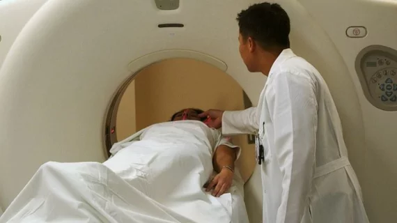
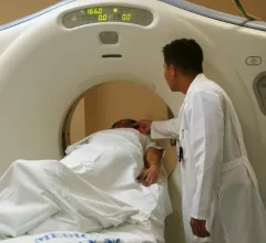
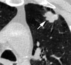
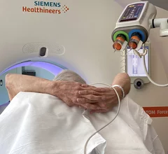
Computed tomography (CT) is a fast and accurate imaging modality often used in emergency settings and trauma imaging. CT scans, with or without (or both) iodinated contrast are frequently used to image the brain, chest, abdomen and pelvis, but also have post-imaging reconstructive capabilities for detailed orthopedic imaging. It is now a standard imaging modality in emergency rooms to quickly assess patients. CT uses a series of X-ray images shot as the gantry rotates around the patient. Computer technology assembles these into into a dataset volume than can be slices on any access, or advanced visualization software can extract specific parts of the anatomy for study. Find more content specific to cardiac CT.

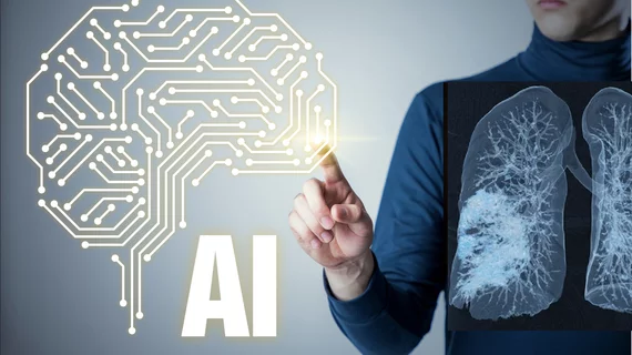
An overview of artificial intelligence (AI) in radiology with Keith Dreyer with the ACR. Images shows a COVID-19 lung CT scan reconstruction from Siemens Healthineers. #AI #radAI #ACR

Ultralow-dose CT aids in diagnosing pneumonia among immunocompromised patients

In a prospective study involving 54 adults, ultralow-dose CT, denoised with deep learning, “substantially” cut radiation exposure while accurately detecting pneumonia. 

Thumbnail

American College of Radiology announces significant expansion of Lung Cancer Screening Registry

The ACR hopes these changes, including the addition of diagnostic performance feedback, will help reduce the number of patients with incidental nodules lost to follow-up each year.

Thumbnail

FDA clears AI-assisted CCTA software that assesses plaques for signs of heart disease

Caristo Diagnostics, an Oxford-based medtech company founded by cardiologists, has gained FDA clearance for its CaRi-Plaque technology.

AI beats standard regression models at predicting lung cancer risk

Not all AI or regression models are the same, nor do they all incorporate the same data when assessing patient risk.

Building a successful CCTA program: Physicians and healthcare executives to share advice

CCTA continues to grow more and more important in the day-to-day treatment of heart patients. Hospitals and health systems that fail to embrace the modality risk falling behind.

Thumbnail

How radiologists can help identify victims of domestic violence

New analysis details patterns radiologists can look for that may signal a patient is experiencing domestic violence. 

AI reduces CT lung cancer screening workload by nearly 80%

And it can do so with almost 100% accuracy as a first reader, according to a new large-scale analysis.

David A. Rosman, MD, MBA, deputy chief, radiology enterprise service, Mass General Brigham, explains details of a recent AJR article that showed imaging outside of hospitals could potentially save billions.

Shift toward imaging outside the hospital could save billions

Harvard’s David A. Rosman, MD, MBA, explains how moving imaging outside of hospitals could save billions of dollars for U.S. healthcare.

Around the web

The cardiac technologies clinicians use for CVD evaluations have changed significantly in recent years, according to a new analysis of CMS data. While some modalities are on the rise, others are being utilized much less than ever before.

The new guidelines were designed to ensure sonographers and other members of the heart team have the information they need to screen patients when appropriate and identify early warnings signs of PH. 

Harvard’s David A. Rosman, MD, MBA, explains how moving imaging outside of hospitals could save billions of dollars for U.S. healthcare.