Nuclear Medicine

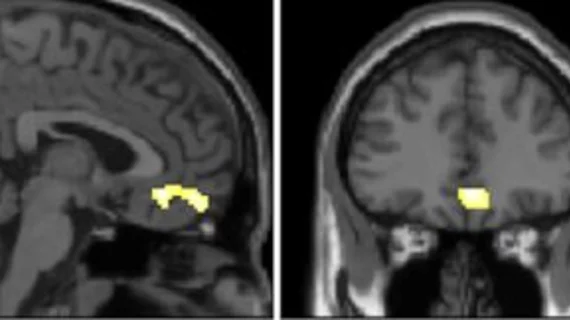
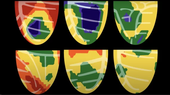
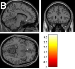
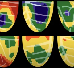
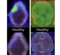
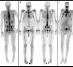
Nuclear medicine (also called molecular imaging) includes positron emission computed tomography (PET) and single photon emission computed tomography (SPECT) imaging. Nuclear imaging is achieved by injecting small amounts of radioactive material (radiopharmaceuticals) into patients before or during their scan. These can use sugars or chemical traits to bond to specific cells. The radioactive material is taken up by cells that consume the sugars. The radiation emitted from inside the body is detected by photon detectors outside the body. Computers take the data to assemble images of the radiation emissions. Nuclear images may appear fuzzy or ghostly rather than the sharper resolution from MRI and CT.  But, it provides metabolic information at a cellular level, showing if there are defects in the function of the heart, areas of very high metabolic activity associated with cancer cells, or areas of inflammation, data not available from other modalities. These noninvasive imaging exams are used to diagnose cancer, heart disease, Alzheimer’s and Parkinson’s disease, bone disorders and other disorders. 

PET method offers new insight into inflammation of the brain

Researchers are hopeful that their technique could improve the diagnosis of conditions associated with neural inflammation. 

Thumbnail

New PET imaging method could improve our understanding of long COVID

Researchers have developed a new radiotracer that targets one of the virus’ spike proteins, allowing them to track it longitudinally.  

FDA approves new prostate cancer imaging agent with extended shelf life

The new product from Telix paves the way for increased access at clinics where prostate PET may not have been available in the past.

Researchers track dopamine receptors to study its impact on cognitive function.

New PET study offers insight into how dopamine drives executive function

These findings could help guide the development of new treatments for psychiatric and neurological disorders.  

HeartSee 4.0 Bracco Diagnostics

Bracco updates HeartSee coronary flow capacity software with new diagnostic features

Clinicians have been using HeartSee to diagnose and treat coronary artery disease since the technology first debuted back in 2018. These latest updates, set to roll out to existing users, are designed to improve diagnostic performance and user access.

lung cancer screening

New PET method combines antibody with radioactive isotope to improve lung cancer detection

By giving providers greater insight into a tumor’s molecular footprint, the method could also reduce the need for invasive biopsies.

ALS drug repurposed for PET imaging

ALS drug repurposed for PET imaging

Researchers radiolabeled the drug so that they could follow its movement and breakdown in the body to help visualize areas of oxidative stress.

cardiac amyloidosis on bone scan

Continuation of 99mTc-PYP shortage prompts need for alternative cardiac amyloidosis imaging

After three years of intermittent shortages of nuclear imaging tracer technetium-99m pyrophosphate, there are no signs of the shortage abating.

Around the web

GE HealthCare designed the new-look Revolution Vibe CT scanner to help hospitals and health systems embrace CCTA and improve overall efficiency.

Clinicians have been using HeartSee to diagnose and treat coronary artery disease since the technology first debuted back in 2018. These latest updates, set to roll out to existing users, are designed to improve diagnostic performance and user access.

The cardiac technologies clinicians use for CVD evaluations have changed significantly in recent years, according to a new analysis of CMS data. While some modalities are on the rise, others are being utilized much less than ever before.