Magnetic Resonance Imaging

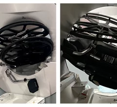
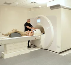
Magnetic resonance imaging (MRI) is the gold standard imaging modality for soft tissues. It produces detail cross-sectional images of soft tissue and bone anatomy, including muscles, tendons, ligaments, brain and organs, without the use of ionizing radiation. In addition to orthopedic imaging, MRI is also used for heart, brain and breast. MRI uses gadolinium contrast in many exams to highlight tissues and blood vessels, which enhances images and offers better diagnostic quality. It can also be used in conjunction with PET scans. How does MRI work? MR creates images by using powerful magnets to polarize hydrogen atoms in water (the body is made of of more than 80% water) so they face in one direction. A radiofrequency pulse is then used to ping these atoms, causing them to wobble, or resonate. The MRI coils detect this and computers can assemble images from the signals. Basic MRI scans will focus on the resonance of fat and water in two different sequences, which highlight and contrast different features in the anatomy.

Thumbnail

Alzheimer's and Parkinson's cause similar cognitive symptoms, but show opposing patterns on MRI

This new information on the pathogenesis of both conditions could be useful for the development of interventions targeted at slowing cognitive decline. 

COVID-19 coronavirus mask smell

Chemical alterations in the brain hint at whether loss of smell will return post-COVID

Researchers have identified what they believe could serve as imaging biomarkers that predict recovery from anosmia after contracting the virus. 

MRI of knee with Osgood-Schlatter Disease

MRI scoring system simplifies diagnosis of common adolescent ortho issue

The condition can worsen with physical activity, which is why an accurate and timely diagnosis is important for healthy growth. 

birth control the pill cardiovascular disease blood clots side effects

Researcher scans herself 75 times to study effects of birth control on brain health

The postdoctoral research fellow hopes her research will provide greater insight into how the use of oral birth control impacts physical and mental health.

Thumbnail

Brain changes related to Huntington's evident on imaging decades before symptoms emerge

Experts hope their findings offer valuable insights that one day lead to improved interventions aimed at preserving brain function in patients with Huntington's. 

Thumbnail

New GBCA uses 60% less gadolinium

Bayer’s latest contrast agent is proving its potential in clinical trials as a promising alternative to agents with higher gadolinium content. 

Thumbnail

Advanced MRI techniques may provide insight into brain damage stemming from youth sports

The study is part of a bigger effort to understand why some children recover better than others following head injuries.

Thumbnail

Basketball players' brains are wired differently, new MRI data suggest

Although some form of neural superiority may not be the driving factor behind an elite athlete’s talent, new evidence shows these individuals have unique structure and connectivity patterns.

Around the web

To fully leverage today's radiology IT systems, standardization is a necessity. Steve Rankin, chief strategy officer for Enlitic, explains how artificial intelligence can help.

RBMA President Peter Moffatt discusses declining reimbursement rates, recruiting challenges and the role of artificial intelligence in transforming the industry.

Deepak Bhatt, MD, director of the Mount Sinai Fuster Heart Hospital and principal investigator of the TRANSFORM trial, explains an emerging technique for cardiac screening: combining coronary CT angiography with artificial intelligence for plaque analysis to create an approach similar to mammography.