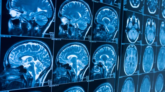
Researchers use MRI scans to develop a growth chart specific to the human brain
A team of researchers have developed a new tool that benchmarks brain development and growth based on thousands of images from MRI scans.
The tool, called BrainChart, was created using imaging from more than 100,000 individuals. Much like the growth tables used in pediatric settings to track children’s height, weight and head circumference, BrainChart can be used to measure brain development against reference standard charts.
“There are no standardized growth charts for brain development like there are for other growth metrics such as height and weight, despite the fact that we know the brain goes through many changes over the human lifespan,” co-senior author Aaron Alexander-Bloch, MD, director of the Brain-Gene-Development Lab within the Department of Child and Adolescent Psychiatry and Behavioral Sciences at Children’s Hospital of Philadelphia, and co-authors wrote. “Our work brings together a huge amount of imaging data that will continue to grow, allowing researchers and eventually clinicians to evaluate brain development against standardized measures.”
To accurately depict the entire life cycle of the human brain for the BrainChart platform, researchers used MRIs from individuals just 115 days after conception to people who were 100 years old at the time of imaging. This resulted in 123,984 MRI scans from 101,457 participants from more than 100 different studies.
Researchers used the images to measure tissue volumes, structural changes and rates of change over an entire lifespan. They quantified these metrics by using centile scores, which they based their developmental charts on. This resulted in the experts uncovering previously unreported neurodevelopmental milestones, such as a period of growth that begins at 17 weeks after conception and ends around the age of 3.
The body of researchers included experts from the Lifespan Brain Institute (LiBI) of Children’s Hospital of Philadelphia (CHOP) and the University of Pennsylvania, as well as a slew of other international team members from all over the world. The researchers noted that although their work is important for the future, it does still need to be validated and refined with more research in order to be able to diagnose neurodevelopmental disorders related to atypical growth.
“Even for traditional anthropometric growth charts (height, weight and BMI), there are still important caveats and nuances concerning their diagnostic interpretation in individual children; similarly, it is expected that considerable further research will be required to validate the clinical diagnostic utility of brain charts,” the authors explained. “However, the current results bode well for future progress towards digital diagnosis of atypical brain structure and development.”
The detailed research is published in Nature.
More on neuroimaging:
MRI scans pinpoint root of lingering concussion symptoms
PET scans spot brain abnormalities in long COVID patients
MRI scans link atypical growth of key brain structure during infancy with autism
'Pandemic brain': PET/MRI images reveal how COVID's impact is felt by non-infected individuals
New advanced PET imaging reveals root of cognitive decline in patients with Alzheimer's

