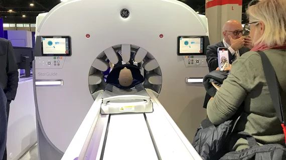
SPECT technique might measure absorbed tissue dose from radiation therapy
Washington University in St. Louis is using a novel low-count quantitative single photon emission computed tomography (LC-QSPECT) technique to measure the concentration of alpha particle radiopharmaceutical therapy activity in the tumor and in radio-sensitive organs of the body.
Patients with cancer that has spread to the bone are sometimes treated with alpha particle radiation therapy that is delivered into, or very close to, the tumor. But, it is unknown whether the radioactive particles are distributed to the surrounding region or to organs elsewhere in the body.
“Understanding the distribution of the drug inside the body helps plan the treatment. Also, one key concern is underdosing, which would not kill the tumor," explained Abhinav Jha, PhD, assistant professor of biomedical engineering at the McKelvey School of Engineering and of Radiology at the School of Medicine’s Mallinckrodt Institute of Radiology (MIR), both at Washington University, in a statement.
Jha and colleagues plan to measure this distribution using a novel imaging method with a four-year $2.2 million grant from the National Institutes of Health (NIH).
Single-photon emission computed tomography (SPECT) nuclear imaging provides a mechanism to estimate regional isotope uptake in lesions and at-risk organs after administration of alpha particle-emitting radiopharmaceutical therapies. But, this estimation is challenging due to the complex emission spectra, a 20 times lower number of detected counts than in conventional SPECT, ands the impact of stray radiation-related noise at these low counts. Conventional approaches that reconstruct the distribution of the isotopes and estimate the uptake from reconstructed images are not accurate at low-count levels, so there is a need for new approaches to quantify the drug in a patient, Jha explained.
To solve for these technical issues, the team is using LC-QSPECT to build a computational framework from which to measure the concentration of the radiopharmaceutical material. Scans with this technology will allow the team to measure the concentration of the radiopharmaceutical activity in the tumor and the various radio-sensitive organs of the body.
Previous research published by the team earlier this year in IEEE Transactions on Radiation and Plasma Sciences,[1] found that a LC-QSPECT method provided reliable measurements of the radionuclide uptake. To validate this method, Jha and his team plan a clinical trial in patients with metastatic prostate cancer that no longer responds to hormone therapy, or is castrate resistant. The team has already done a computational clinical trial in a simulated patient population. The results showed the method was yielding highly accurate and precise values of radionuclide organ uptake.
“Our eventual goal is clinical translation of this method so that it can benefit patients being treated with these therapies.” Jha explained. “There is much excitement surrounding this use of SPECT imaging for therapy. We are delighted to have the opportunity to contribute to this space.”
The team also published on this method in the June 1, issue of Journal of Nuclear Medicine.[2]

