CT findings linked to heightened risk of pancreatic cancer recurrence
Findings related to soft tissue in surveillance imaging CT scans may indicate that patients in remission from pancreatic cancer could be at an increased risk of recurrence.
In 2023, the Society of Abdominal Radiology (SAR) released its Pancreatic Ductal Adenocarcinoma (PDAC) Disease-Focused Panel consensus statement regarding follow-up imaging of patients who have had their cancer surgically resected. A new paper in the American Journal of Roentgenology reviews these recommendations, comparing them to a set of 126 patients who underwent Whipple surgery for PDAC between 2009 and 2014.
“Given that surveillance imaging entails performing comparisons with prior examinations and accounting for interval changes in findings, the features described in the SAR PDAC consensus statement should be evaluated across serial surveillance CT examinations, rather than assessed solely on the first postoperative CT,” corresponding author Tae-Hyung Kim, MD, from the Department of Radiology at Memorial Sloan Kettering Cancer Center in New York, and colleagues suggested. “Moreover, since the consensus statement was derived from expert opinion, further clinical testing is needed.”
The team’s analysis consisted of having three radiologists independently review pre- and post-operative contrast enhanced CT scans of the patients. The scans were completed within two years of the patients’ surgical resection, with findings compared to the features included in SAR’s consensus statement relating to surgical bed stranding, surgical bed soft tissue, vessel encasement, main pancreatic duct dilatation and ascites.
Local recurrence was observed in 81 out of 126 patients. Findings related to main pancreatic duct dilation had the highest agreement among readers on both pre- and post-op imaging, while stranding and soft tissue morphology yielded the lowest.
On recurrence examinations, the following was observed:
New or increased stranding in 27%-77% of patients.
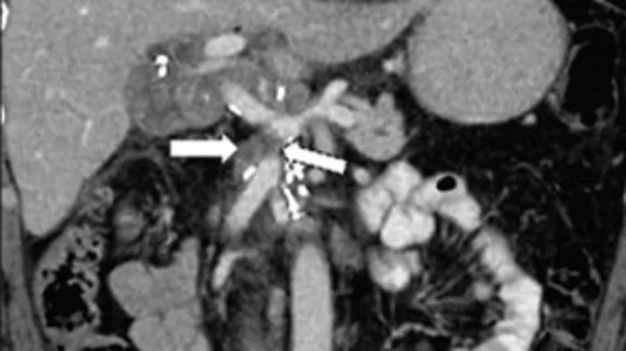
New or increased soft tissue in 80%-86%.
Soft tissue with vessel encasement and luminal narrowing in 36%-59%.
New or increased main pancreatic duct dilatation in 25%-26%.
New or increased ascites in 20%-23%.
On baseline imaging, alterations in soft tissue were the greatest predictor of local recurrence across all three readers.
The findings support SAR’s consensus statement and offer further guidance on imaging features that warrant added concern from radiologists interpreting the exams of PDAC patients, the group suggested.

