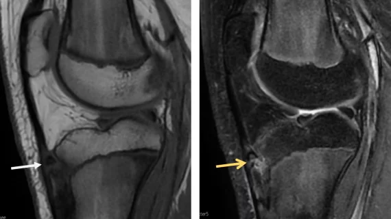
MRI scoring system simplifies diagnosis of common adolescent ortho issue
Experts have developed a new MRI scoring system for a common orthopedic condition that ails adolescents.
Osgood-Schlatter Disease (OSD) causes pain and swelling just below the knee where the patellar tendon attaches to the tibia. It is commonly diagnosed during adolescent growth spurts and is easily treated with rest and ice. However, the condition can worsen with physical activity, which is why an accurate and timely diagnosis is important for healthy growth.
Clinicians often diagnose OSD without the use of imaging, but in cases of uncertainty, MRI and ultrasound can be used to gain a more detailed depiction of the condition’s severity and, therefore, provide a more accurate diagnosis. However, there are no established diagnostic criteria for assessing OSD on imaging, authors of a new paper in the European Journal of Radiology explain.
“A systematic approach to rating MR images from adolescents with OSD is currently lacking,” M.S. Rathleff, with the Department of Occupational Therapy and Physiotherapy at Aalborg University Hospital in Denmark, and colleagues note. “This hampers our understanding of the tissue characteristics associated with OSD and our ability to detect changes across time.”
The new 18-item scoring system was developed by a group of musculoskeletal experts and reviewed by 2 radiologists. The list describes different imaging characteristics of the patellar tendon, infrapatellar bursa, cartilage, and the tibial epiphysis, metaphysis and tibial tuberosity. Its efficacy was tested on a group of children with OSD using a 1.5T MRI scanner.
There was agreement among readers that most of the chosen features could help reliably diagnose OSD, with measurements of patellar height and patellar tendon width being the greatest predictors. Intra-rater reliability for superficial bursa homogeneity and inter-rater reliability for signal degree in the patellar tendon, tibial epiphysis and tibial tuberosity morphology ratio were the least reliable metrics.
“This novel and reliable semi-quantitative scoring and morphometric system for MRI allows for a comprehensive assessment of a range of items that may be relevant for reporting and quantifying tissue abnormalities in adolescents with OSD,” the authors suggest.
The team acknowledges that imaging may not always be necessary to diagnose OSD, but points to recent data indicating that nearly half of providers would prefer to routinely utilize imaging to assess the condition. MRI will not always be the first-line modality for OSD but could serve as a reliable tool for more complex cases, the group signals.
Learn more about the findings here.

