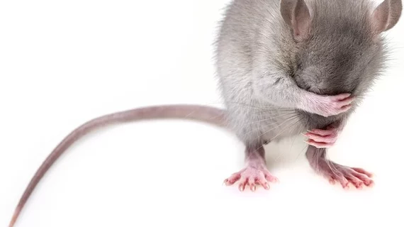
Rodent brains retain gadolinium after repeated administration of GBCA a year after injection
Researchers from the Guerbet Group and the University of Münster in Germany recently found that 75 percent of the total gadolinium found in the cerebellum of a rat's brain after the injection period of the linear contrast agent gadodiamide was still retained after one year.
The study aimed to compare how the brain's cerebellum eliminates gadolinium after repeated injections of the contrast agents gadodiamide (a linear contrast agent) or gadoterate (a macrocyclic contrast agent). Findings were published online on May 22 in Radiology.
"After repeated administration of gadodiamide, a large portion of gadolinium was retained in the brain, with binding of soluble gadolinium to macromolecules," wrote lead author Philippe Robert, PhD, and colleagues. "After repeated injection of gadoterate, only traces of the intact chelated gadolinium were observed with time-dependent clearance."
To analyze gadolinium brain retention in kinetic and molecular forms, Robert and his team analyzed 140 nine-week-old rats, all of which received five doses of 2.4 mmol gadolinium per kilogram of body weight over five weeks.
The rats were then monitored for one year with T1-weighted MRI and sacrificed at various time points post injection for researchers to accumulate brain and plasma samples from the rats' cerebellums, the researchers wrote.
Despite their findings, however, the researchers prompted for additional research to conducted in order to clearly characterize the nature of the macromolecules that the contrast agent binds itself to.
Related MRI Contrast Agent Safety Content:
A deep dive into gadolinium-based adverse reactions
Allergic reactions to iodinated CT contrast increase likelihood of sensitivity to GBCAs
Researchers detail data on gadolinium-related adverse reactions
Radiologists must take a data-driven approach to discuss gadolinium, mitigate liability risk
Radiologists see potential to reduce GBCA administration with new synthetic MRI technique
Gadolinium-based contrast agents are safe, even at higher doses, new research suggests
Gadolinium debate rages on, with radiologist questioning recent GBCA liability guidance
ACR committee proposes new term for symptoms associated with gadolinium exposure
Closing the knowledge gap on gadolinium retention risks
Radiologists find direct evidence linking gadolinium-based contrast agent to higher retention rates
AI software that eliminates need for gadolinium contrast during imaging exams wins patent
Research may offer new method to detect GBCA on MRI
Radiology, other multispecialty groups urge caution with GBCAs during interventional pain procedures
Cardiac MRI contrast agents are low-risk and safe for ‘overwhelming’ majority of patients
Health orgs publish special report about gadolinium retention, GBCA use in imaging
Rodent brains retain gadolinium after repeated administration of GBCA a year after injection
Advanced MRI mapping spots traces of gadolinium in the brain invisible during conventional scanning
Radiologists should keep patients’ best interests in mind to mitigate gadolinium liability risk

