Molecular Imaging

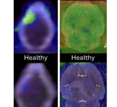
Molecular imaging (also called nuclear medicine or nuclear imaging) can image the function of cells inside the body at the molecular level. This includes the imaging modalities of positron emission computed tomography (PET) and single photon emission computed tomography (SPECT) imaging. How does PET and SPECT imaging work? Small amounts of radioactive material (radiopharmaceuticals) injected into a patient. These can use sugars or chemical traits to bond to specific cells. The radioactive material is taken up by cells that consume the sugars. The radiation emitted from inside the body is detected by photon detectors outside the body. Computers take the data to assemble images of the radiation emissions. Nuclear images may appear fuzzy or ghostly rather than the sharper resolution from MRI and CT.  But, it provides metabolic information at a cellular level, showing if there are defects in the function of the heart, areas of very high metabolic activity associated with cancer cells, or areas of inflammation, data not available from other modalities. These noninvasive imaging exams are used to diagnose cancer, heart disease, Alzheimer’s and Parkinson’s disease, bone disorders and other disorders. 

Thumbnail

Repurposed radiotracer lights up treatment-resistant tumors 'like a Christmas tree'

Use of the new tracer could improve outcomes by enabling providers to adjust treatment regimens much earlier than they have been able to previously. 

Researchers working to develop portable PET scanner

Researchers receive grant worth millions to develop portable PET scanner

Experts are hopeful that the portable scanner could improve access to neuroimaging used in the diagnosis and monitoring of Alzheimer's disease.

Why is the US still dependent on foreign medical isotope production?

The U.S currently does not have any large-scale commercial production of the isotope Mo-99 used for nuclear imaging. It is critical to more 40,000 nuclear imaging exams each day, representing about 80% of all diagnostic molecular imaging procedures.  

Downed high-flux reactor could restart sooner than anticipated

The announcement will come as welcome news, as nuclear medicine departments in numerous regions have been grappling with shortages of Mo-99 and Tc-99m.

maya kowalski case

Some nuc med departments are already feeling the strain of the isotope shortage

"This can't drag on. Patients needing cancer treatment can’t wait two months for an accurate diagnosis.” 

NorthStar opens CDMO facility to help develop and distribute radioisotopes.

NorthStar opens facility to assist with development and distribution of vital radioisotopes

The facility will provide much needed developmental and manufacturing support for pharmaceutical companies that produce and distribute medical isotopes.

Gila monster saliva helps docs detect insulinomas.

Lizard saliva could be key to spotting elusive pancreatic tumors

By binding the substance to a radiotracer, experts were able to improve the accuracy of identifying these tumors by as much as 30% compared to the standard of care.

Thumbnail

New details emerge on looming medical isotope shortage

The supply of Mo-99 and Tc-99m—the most commonly utilized medical isotopes in the world—could be cut by as much as 40% until the reactor in the Netherlands is up and running again.

Around the web

The new technology shows early potential to make a significant impact on imaging workflows and patient care. 

Richard Heller III, MD, RSNA board member and senior VP of policy at Radiology Partners, offers an overview of policies in Congress that are directly impacting imaging.
 

The two companies aim to improve patient access to high-quality MRI scans by combining their artificial intelligence capabilities.