Magnetic Resonance Imaging

Magnetic resonance imaging (MRI) is the gold standard imaging modality for soft tissues. It produces detail cross-sectional images of soft tissue and bone anatomy, including muscles, tendons, ligaments, brain and organs, without the use of ionizing radiation. In addition to orthopedic imaging, MRI is also used for heart, brain and breast. MRI uses gadolinium contrast in many exams to highlight tissues and blood vessels, which enhances images and offers better diagnostic quality. It can also be used in conjunction with PET scans. How does MRI work? MR creates images by using powerful magnets to polarize hydrogen atoms in water (the body is made of of more than 80% water) so they face in one direction. A radiofrequency pulse is then used to ping these atoms, causing them to wobble, or resonate. The MRI coils detect this and computers can assemble images from the signals. Basic MRI scans will focus on the resonance of fat and water in two different sequences, which highlight and contrast different features in the anatomy.

Thumbnail

MRI guidance significantly reduces radiotherapy side effects compared to CT

A recent comparison of the two modalities’ utility for guiding radiation therapy for prostate cancer shows numerous benefits for MRI over CT.

Thumbnail

Socioeconomic disadvantage during childhood linked to altered brain structure later in life, MRIs show

New MRI data highlights associations between being born into disadvantaged communities and decreased volume of multiple areas in the brain. What’s more, these findings hold true despite educational attainment and late-life income.  

Thumbnail

Deep learning reconstruction cuts prostate MRI acquisition time

And the shorter scan time does not come at the expense of image quality.

Thumbnail

The Mediterranean diet may slow brain aging, MRI study shows

Following the Mediterranean diet, especially one rich with plant-based foods, could have significant benefits for cognitive health. 

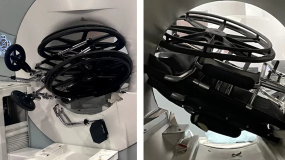
MRI accident prompts questions on safety

CMS to make proposed MRI safety codes official

The new code family was created to "capture the physician work involving patients with implanted medical devices that require access to MR diagnostic procedures."

differentiating between malignant and vaccine-related lymphadenopathy

7 COVID vaccine complications: The radiologist's role in identifying adverse reactions

Although serious adverse events related to COVID-19 vaccines are rare, some are life-threatening. It is important that radiologists are aware of how these complications present.

AI prostate lesion segmentation algorithm

AI tumor volume estimations could improve prostate cancer treatment strategies

More detailed estimations of lesion size could give providers greater insight into how to best manage a patient’s treatment plan. 

Rads' lumbar MRI reads more accurate when they have access to patients' self-reported symptoms

Rads are more accurate when they have access to patients' self-reported symptoms

This is especially true for lumbar MRIs, as these exams often show abnormalities without correlating symptoms, making it difficult to distinguish between incidental findings and the actual source of patients’ issues.

Around the web

The nuclear imaging isotope shortage of molybdenum-99 may be over now that the sidelined reactor is restarting. ASNC's president says PET and new SPECT technologies helped cardiac imaging labs better weather the storm.

CMS has more than doubled the CCTA payment rate from $175 to $357.13. The move, expected to have a significant impact on the utilization of cardiac CT, received immediate praise from imaging specialists.

The newly cleared offering, AutoChamber, was designed with opportunistic screening in mind. It can evaluate many different kinds of CT images, including those originally gathered to screen patients for lung cancer. 

Trimed Popup
Trimed Popup