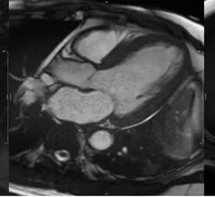
Magnetic Resonance Imaging

Cardiac magnetic resonance imaging MRI is used as both a functional and anatomical cardiac imaging test. It offers excellent soft tissue detail and the ability to quantify cardiac function. MRI scans can be performed with or without gadolinium contrast depending on what information is needed. Unlike computed tomography (CT), MRI does not use X-ray radiation, but patients with metal implants may have contraindications for MRI use because MR will heat up most metal objects. MRI exams usually take much longer than CT scans. How does MRI work? MR creates images by using powerful magnets to polarize hydrogen atoms in water (the body is made of of more than 80% water) so they face in one direction. A radiofrequency pulse is then used to ping these atoms, causing them to wobble, or resonate. The MRI coils detect this and computers can assemble images from the signals. Basic MRI scans will focus on the resonance of fat and water in two different sequences, which highlight and contrast different features in the anatomy.
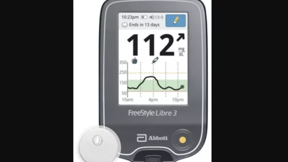
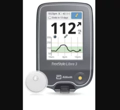
Abbott’s FreeStyle Libre 3 integrated continuous glucose monitoring (iCGM) system

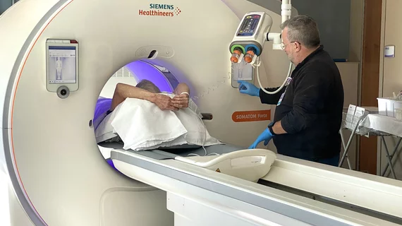
Abbott CGM sensors can now be worn during common imaging procedures

Sensors from the FreeStyle Libre 2 and Libre 3 continuous glucose monitoring systems can now be worn during X-rays, CT scans and MRI scans. The news represents a shift in policy from the FDA, one that came after the agency reviewed extensive testing data.

The American Society of Echocardiography (ASE) has shared new recommendations for performing medical imaging exams on heart failure patients with surgically implanted left ventricular assist devices (LVADs) and temporary mechanical circulatory support (TMCS) devices. The guideline, published in full in the Journal of the American Society of Echocardiography, represents ASE’s first update on the topic since 2015.[1]

ASE shares new guideline on imaging patients with implanted devices

The new guideline details the best imaging strategies for a variety of clinical scenarios. 

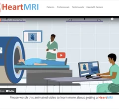
The Society of Cardiovascular Magnetic Resonance (SCMR) recently launched HeartMRI.org, which provides includes a general information and videos for patients and referring physicians.

SCMR launches cardiac MRI educational video to assist referring physicians 

The Society of Cardiovascular Magnetic Resonance launched a new website aimed at patients and referring physicians that includes pamphlets and videos on cardiac MRI. 

Ron Blankstein, MD, FACC, FASNC, MSCCT, FASPC, associate director, cardiovascular imaging program, director, cardiac computed tomography, Brigham and Women’s Hospital, and a professor of medicine and radiology, Harvard Medical School, explains a few of the recent advancements in cardiac MRI, nuclear imaging and computed tomography.

Exploring major breakthroughs in cardiac imaging

Ron Blankstein, MD, director of cardiac computed tomography for Brigham and Women’s Hospital, details recent developments in cardiac MRI, nuclear imaging and CT.

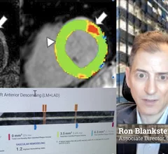
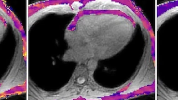
CMR of a patient showing evidence of myocardial hypertrophy

MRI results help cardiologists predict heart failure risk

"This breakthrough suggests that heart MRI could potentially replace invasive diagnostic tests," one specialist said. 

Researchers have developed and validated a new artificial intelligence (AI) model capable of producing four-chamber cardiac MR (CMR) images in seconds, publishing their findings in European Radiology Experimental.

AI creates accurate 4D heart scans in seconds

New data out of England provides cardiologists and radiologists with another reminder of AI's potential to transform healthcare. 

Thumbnail

New imaging protocols proposed to curb rise of cardiovascular infections

Eleven medical societies have signed on to a consensus statement aimed at standardizing imaging for suspected cardiovascular infections.

Kate Hanneman, MD, University Of Toronto, explains why vendors and hospitals are increasingly discussing lowing their carbon footprint by starting with radiology. 

What does radiology have to do with climate change?

Kate Hanneman, MD, explains why many vendors and hospitals want to lower radiology's impact on the environment. "Taking steps to reduce the carbon footprint in healthcare isn’t just an opportunity," she said. "It’s also a responsibility."

Around the web

The nuclear imaging isotope shortage of molybdenum-99 may be over now that the sidelined reactor is restarting. ASNC's president says PET and new SPECT technologies helped cardiac imaging labs better weather the storm.

CMS has more than doubled the CCTA payment rate from $175 to $357.13. The move, expected to have a significant impact on the utilization of cardiac CT, received immediate praise from imaging specialists.

The newly cleared offering, AutoChamber, was designed with opportunistic screening in mind. It can evaluate many different kinds of CT images, including those originally gathered to screen patients for lung cancer. 

Trimed Popup
Trimed Popup