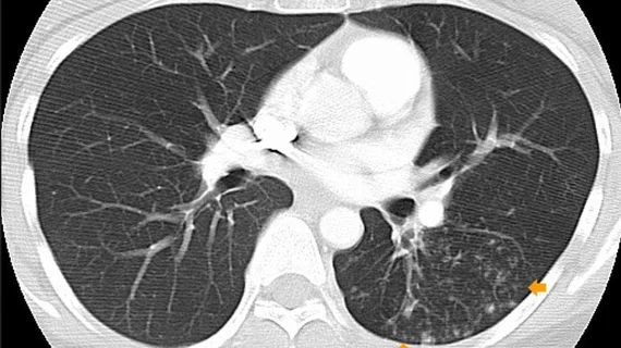
Study urges radiologists to report CAC findings on all chest CTs, regardless of clinical indication
Reporting findings of coronary artery calcium (CAC) identified on any computed tomography scan, regardless of their clinical indication, could lead to earlier interventions for patients who were previously unaware of their risks.
A new study out of Toronto General Hospital that was published in the American Journal of Roentgenology compared the findings of patients’ non-gated CT scans (contrast-enhanced and without contrast) to that of their cardiac CT CAC scores and found that both were indicative of cardiac event outcomes. Although current guidelines recommend radiologists evaluate CAC on all non-gated, non-contrast chest CT scans, the authors of the study note that these guidelines are not consistently followed, nor do they include reporting on findings visualized on contrast-enhanced scans.
The study included an analysis of 260 patients who underwent both non-gated chest CT scans (116 contrast-enhanced and 144 without contrast) and a cardiac calcium score CT in a 12-month time frame. A cardiothoracic radiologist visually assessed coronary calcium on all scans and rated its presence and severity as absent, mild, moderate or severe. Experts recorded major cardiac event outcomes over a 6-year period.
All scans yielded a sensitivity for CAC of 83% and 90%, and a specificity of 100%. Each scanning method was found to be accurate in predicting major cardiac event outcomes, and interobserver agreement between contrast-enhanced and non-contrast chest CT scans was recorded as “excellent.”
“Visual ordinal CAC assessment on both contrast-enhanced and non-contrast chest CT has high diagnostic performance, prognostic utility, and interobserver agreement,” corresponding author Kate Hanneman, MD, MPH, from the Department of Medical Imaging at Toronto General Hospital, and co-authors concluded. “Routine reporting of CAC on all chest CT examinations regardless of clinical indication and contrast material administration could identify a large number of patients with previously unknown CAC who might benefit from preventive treatment.”
Related cardiac imaging content:
PHOTO GALLERY: Duly Health adopts outpatient cardiac CT as a standard of care
Risks of stroke and heart attack increase with larger thoracic aortic diameter, research shows
AI tool detects CVD on cardiac MRI in 20 seconds with high precision

