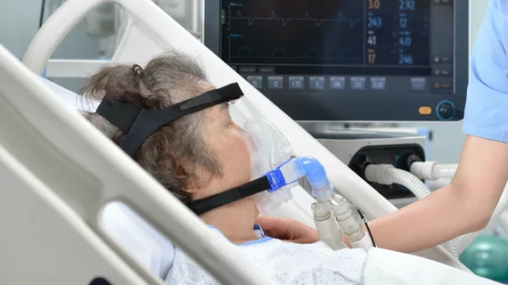
More than 50% of patients hospitalized with COVID-19 retain heart damage a month after discharge
More than 50% of patients hospitalized with severe COVID-19 and high levels of a protein known as troponin show heart damage one month after discharge, according to a multicenter analysis published Thursday.
University College London researchers used cardiac MRI to analyze 148 patients with the virus, 32% of whom needed ventilation. Overall, 54% had heart muscle injuries, including inflammation, tissue scarring, restricted blood blow, or a combination of all three, the group reported Feb. 18 in the European Heart Journal.
The researchers say their study is the largest of its kind and noted the patient data may help spot various injury patterns, potentially enabling more accurate diagnoses and treatment.
“Importantly, the pattern of damage to the heart was variable, suggesting that the heart is at risk of different types of injury,” Marianna Fontana, MD, PhD, a cardiology professor at the U.K.-based institution, said in a statement. “In the most severe cases, there are concerns that this injury may increase the risks of heart failure in the future, but more work is needed to investigate this further.
To reach their conclusions, Fontana et al. imaged patients treated across six acute hospitals in London at the beginning of the pandemic and 40 healthy volunteers. Each participant admitted to the ICU had elevated troponin levels suggestive of heart damage, with MRI scans gathered at least one month after they were discharged.
Of the 148 patients, 80 showed heart muscle injuries. Inflammation was responsible for such problems in 26% of patients, ischemic heart disease in 22%, and for 6% of participants both contributed to their diagnosis. On the other hand, 89% of the group showed normal left ventricle function, indicating many retained regulated oxygenation throughout the body.
While the results say “nothing” about those who don’t require COVID-19-related hospitalization, Fontana says the research will help direct care to people most in need.
“The findings indicate potential ways to identify patients at higher or lower risk and suggest potential strategies that may improve outcomes,” Fontana added. “More work is needed, and MRI scans of the heart have shown how useful it is in investigating patients with troponin elevation.”

