COVID long haulers have enlarged brain stems
Experts recently found imaging similarities in the brains of people who suffer from chronic fatigue syndrome (CFS) and long COVID.
Using a powerful up and coming imaging tool—7T MRI—researchers noted that individuals who suffer from CFS (also known as myalgic encephalomyelitis ) and long COVID have something in common on imaging—enlarged brain stems. In comparison to a cohort of individuals who were unaffected by either condition, the difference in brain stem sizes was found to be significant, according to a paper published recently in Frontiers in Neuroscience.
One of the paper’s lead authors, Kiran Thapaliya, with the Center for Advanced Imaging at the University of Queensland in Australia, suggested that the similarities in brain stem volumes between individuals with CFS and COVID long haulers could be a clue as to why those suffering from long COVID so often report feeling chronic, severe fatigue. The experts also found links between reduced midbrain volumes and increased respiratory issues in both CFS and long COVID patients.
“Therefore, brainstem dysfunction in ME/CFS and long COVID patients could contribute to their neurological, cardiorespiratory symptoms, and movement disorder,” the group suggested.
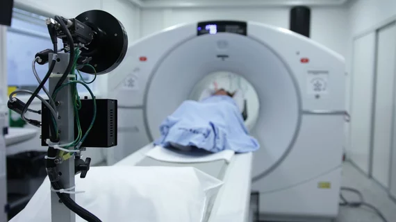
The study was small, consisting of just 10 CFS patients, eight COVID long haulers and 10 healthy controls; however, experts involved in this latest research indicated that their work is significant not just because of the new data it provided, but also because of their use of 7T MR imaging, as it enabled them to visualize areas of the brain that less powerful MRI equipment cannot.
“We primarily used the 7T MRI to research the brainstem and its sub regions as it helps to resolve brain structures more precisely,” the group explained.
In comparison to lower field MRI systems, such as 1.5T and 3T, there are very few 7T systems in clinical use across the world. Australia has just two of them.
To view the full study, click here.

