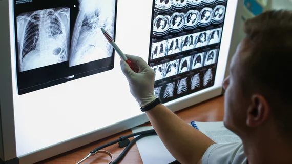
Intrathoracic complications in COVID patients: Incidence, associations and outcomes
Intrathoracic complications are frequently seen in imaging of patients with COVID and new research now links specific findings to ICU admission, need for intubation and length of hospital stay.
Experts recently analyzed the chest imaging of more than 900 COVID patients to look for intrathoracic complications such as pneumothorax, pneumomediastinum, pneumopericardium, lobar collapse, pleural effusion and pneumatocele formation. Through their efforts, they found that 1 in 5 patients diagnosed with the respiratory virus exhibit complications on imaging.
“The most common complication of COVID-19 pneumonia is acute respiratory distress syndrome (ARDS). However, there are additional intrathoracic complications [that] may occur in conjunction with or separate from ARDS,” corresponding author Joanna G. Escalon, MD, from the department of radiology at Weill Cornell Medicine in New York, and co-authors explained. “These are important as they may affect management and can potentially increase morbidity and mortality.”
The study examined 3,836 chest radiographs and 105 CT scans. While 20% of the images uncovered complications, pleural effusion was the most frequently observed, appearing in 17% of exams. This was followed by pneumatoceles (10%) and pneumothorax (3%).
“This adds to our understanding of the pathophysiology of COVID-19 and has several implications for patient care,” the authors wrote.
The complications were seen most often in older patients with a history of smoking, hypertension, CAD, heart failure and high initial CXR severity scores (4 or higher). The findings were linked to an 11-fold increase to a patient’s risk of ICU admission and need for intubation, as well as a 50% decrease in successful extubation. These individuals also underwent longer hospital stays (median 13 vs. 5 days with no complications), but interestingly their adverse findings were not indicative of increased mortality in COVID patients.
“The ability to predict who may need a higher level of care may aid in clinical decision-making such as degree of patient monitoring (i.e., remote home monitoring versus admission),” the experts shared. “Furthermore, prognostic information gleaned from imaging can inform conversations between physicians, patients and their families, potentially alleviating patient and family anxiety and facilitating goals of care discussions.”
The researchers noted the association between image findings and outcomes further underscores the importance of the radiologist’s role on a multidisciplinary COVID team, suggesting that they should be routinely and actively involved in care discussions.
The detailed research can be viewed in Clinical Imaging.
More COVID content:
These ultrasound features distinguish between COVID vaccine-related and malignant adenopathy
'Pandemic brain': PET/MRI images reveal how COVID's impact is felt by non-infected individuals
Cardiac MRI scans offer new insight into COVID vaccine-related myocarditis
Convolutional neural network pipeline has 100% accuracy distinguishing between COVID and pneumonia
Specific chest CT findings linked with increased mortality in COVID patients
Myocarditis, arrhythmias and more: An ACC update on what cardiologists know about long COVID-19
Athletes with COVID-19 may require heart MRI screening for myocarditis, new data suggest
4 cardiac arrhythmias associated with COVID-19
What we know about COVID-19 and cardiogenic shock
Mild COVID-19 infections not associated with long-term risk of heart damage
The pandemic’s toll: 55 long-term side effects of COVID-19
4 key takeaways from an updated look at vaccine-related myocarditis in the U.S.
Most young people with vaccine-related myocarditis recover quickly

