
Experts describe how long-term lung abnormalities of COVID patients present on imaging
Many patients who contracted COVID pneumonia at some point during the last three years display lung abnormalities on imaging for a year or longer after recovering from their initial infection.
What’s worse is that some of these patients remain symptomatic due to the sustained damage in their lungs. This and other revelations were reported on Aug. 20 in a paper published in Radiology, where experts shared that their findings are reminiscent of the abnormalities described following the 2002-2003 SARS epidemic.
In the new paper, corresponding author Jeffrey P. Kanne, of the University of Wisconsin School of Medicine and Public Health, and co-authors reinforced the importance of imaging’s role in finding the root cause(s) of chronic respiratory symptoms in survivors, suggesting that it could help uncover new disease management strategies.
“As the pool of individuals who have suffered one or more episodes of COVID-19 rapidly grows, the proportion of the population with long-term symptoms and chronic lung findings of the disease increases,” the authors wrote. “While the causes of persistent symptoms are currently not well understood, the growing radiology literature on chronic lung findings in COVID-19 may eventually facilitate understanding of long-term respiratory issues in afflicted individuals.”
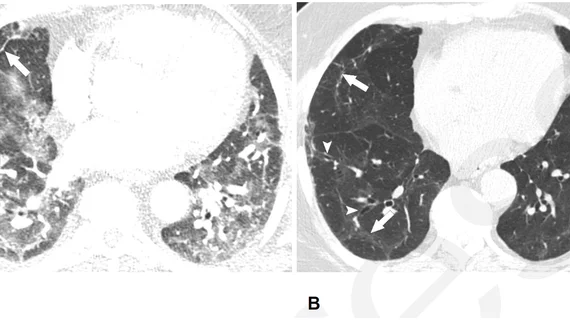
Kanne and colleagues analyzed multiple studies and meta-analyses describing post-COVID lung abnormalities and found that up to one-third of patients hospitalized with COVID-19 pneumonia have abnormalities at chest CT 12 months after infection. These abnormalities manifest on imaging a variety of ways, but most often range from residual parenchymal bands to fibrosis, air trapping and bronchiectasis. The authors also noted that a very small number of patients remain at an increased risk of venothromboembolic disease after their initial infection.
The authors acknowledged that while their meta-analysis does offer some clarity in what is commonly encountered with this cohort of patients, their findings are inevitably impacted by the variability of the studies included.
They went on to suggest several recommendations geared toward the imaging management of these patients, including inspiratory thin-section chest CT with expiratory imaging as indicated, chest CT pulmonary angiography and XeMRI, depending on each patient’s individual signs and symptoms.
For more information, click here.

In addition to her background in journalism, Hannah also has patient-facing experience in clinical settings, having spent more than 12 years working as a registered rad tech. She began covering the medical imaging industry for Innovate Healthcare in 2021.