How do Omicron and Delta compare on chest imaging?
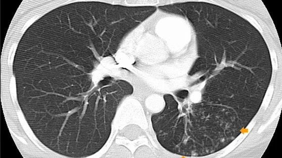
The Omicron variant of COVID revealed itself to be less severe than the Delta variant on imaging, according to new comparative analysis of chest CTs acquired during both viral waves.
The retrospective study consisted of 106 patients—66 with the Delta variant and 40 with Omicron—who underwent pulmonary CT angiography within 7 days of hospitalization. Researchers found that 37% of patients from the Omicron group had scans that were considered normal compared to 15% in the Delta group.
Experts involved in the study explained that patients with the Omicron variant are 50% less likely to be hospitalized or come down with severe illness in comparison to those with the Delta variant. Additionally, those who are vaccinated against COVID have even less of a chance of hospitalization. But statistics regarding the patients who do end up hospitalized with Omicron are limited.
“Establishing intrinsic differences in variant virulence, as opposed to reductions in disease severity over time due to population immunity from vaccination or prior infection, is challenging,” corresponding author Maria T. Tsakok, of Oxford University Hospitals NHS Foundation Trust, and co-authors shared. “Studies have evaluated the CT imaging characteristics or ‘signature’ of SARS-COV-2 infection, but studies evaluating the lung imaging findings of Omicron infection versus non-Omicron variants remain lacking.”
Additional insights gained from the experts’ research include lower CT severity scores (7.2) and increased incidence of bronchial wall thickening visualized in the Omicron group. Regarding vaccination status, those who were vaccinated also recorded lower CT severity scores. These findings were consistent, regardless of the interpreting radiologist’s experience level.
“Our results suggest that infection with Omicron variant is intrinsically less severe than Delta variant infection, as evidenced by the significant difference in severity amongst the unvaccinated during our study period,” Tsakok and colleagues said.
The authors acknowledged that their study did have limitations, such as the subjectivity of identifying bronchial wall thickening, but overall believe that their work validates previous research citing less severe disease and better outcomes with Omicron.
View the full research in Radiology.
Reference:
Chest CT and Hospital Outcomes in Patients with Omicron Compared with Delta Variant SARS-CoV-2 Infection. Maria T. Tsakok, Robert A. Watson, Shyamal J. Saujani, Mark Kong, Cheng Xie, Heiko Peschl, Louise Wing, Fiona K. MacLeod, Brian Shine, Nick P. Talbot, Rachel E. Benamore, David W. Eyre, and Fergus Gleeson. Radiology.
More on COVID in imaging:
Multimodality imaging turns up serial thromboses following AstraZeneca COVID vaccination
New research compares COVID pneumonia in vaccinated vs unvaccinated patients
New imaging technique detects post-COVID lung abnormalities
Reactive lymphadenopathy slower to resolve after Moderna COVID vaccination
Lung abnormalities completely resolve for majority of COVID pneumonia patients

