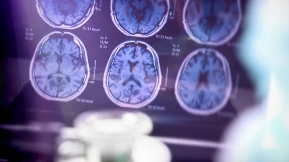
PET scans spot brain abnormalities in long COVID patients
After examining PET images of patients with suspected long COVID, a study published in the European Journal of Nuclear Medicine and Molecular Imaging identified hypometabolic patterns in nearly half of all patients.
Each of the 143 patients was experiencing neurological symptoms at the time of the 18F-FDG PET scan, such as fatigue, pain, memory or cognitive complaints and insomnia. On average, the scans were conducted 10.9 months after the onset of these symptoms.
While 53% of the scans were normal, 21% were classified as mildly to moderately affected according to previously reported COVID hypometabolic patterns and 26% were classified as severely affected. Experienced nuclear physicians classified each of the scans.
“This pattern can be found at an individual level and helps to objectively assess metabolic abnormalities in patients with suspected neurological long COVID,” wrote lead author Antone Verger, of the Department of Nuclear Medicine at the Université de Lorraine in Nancy, France, and co-authors.
The study builds on previous work which had initially identified hypometabolic patterns in long COVID patients and had called for additional, multicenter studies with a larger number of patients. The results appear to validate the ability of nuclear physicians to identify brain abnormalities in long COVID patients, and indicate a possible role for the continued use of PET scans to assess neurological damage both on an individual and a group level.
“Nuclear neurology can be helpful in routine practice for selected patients to identify the possible brain impairment associated with long COVID,” Verger wrote. "The proposed PET metabolic pattern is easily identified upon visual interpretation for approximately one half of patients with suspected neurological long COVID."
With long COVID—defined as the persistence or recurrence of symptoms three months after an initial infection—affecting approximately 10-15% of patients, the study notes that exams can serve a dual purpose. Not only can they reassure symptomatic patients that end up with normal scans, but they can also contribute to social and medical recognition of a patient’s situation when scans show an objective abnormality.
“In such cases of brain involvement observed with 18F-FDG PET, adapted follow-up and medical care should be proposed,” Verger wrote.
More on imaging of COVID:
MRI scans show COVID's 'significant' impact on the brain
Intrathoracic complications in COVID patients: Incidence, associations and outcomes
Imaging suggests blood clots are more common in COVID than pneumonia
Boston researchers hope PET-MRI brain scans will shed light on 'long-COVID' symptoms
