Computed Tomography

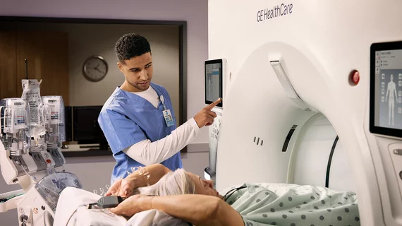
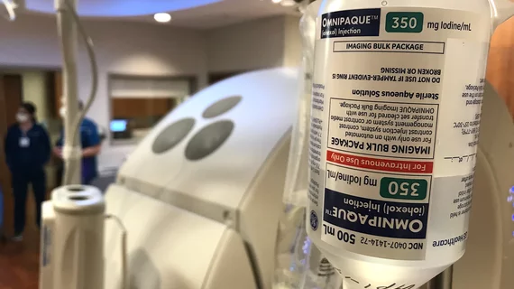
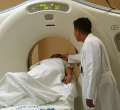
Computed tomography (CT) is a fast and accurate imaging modality often used in emergency settings and trauma imaging. CT scans, with or without (or both) iodinated contrast are frequently used to image the brain, chest, abdomen and pelvis, but also have post-imaging reconstructive capabilities for detailed orthopedic imaging. It is now a standard imaging modality in emergency rooms to quickly assess patients. CT uses a series of X-ray images shot as the gantry rotates around the patient. Computer technology assembles these into into a dataset volume than can be slices on any access, or advanced visualization software can extract specific parts of the anatomy for study. Find more content specific to cardiac CT.

Ischemic stroke shown in CT scans. Image courtesy of RSNA

New algorithm is twice as accurate at predicting stroke timing compared to the standard of care

Determining stroke onset is critical for management, as there is a small window of time for initiating treatment that can inhibit damage.

New protocol addresses contrast allergies in patients with suspected stroke

In patients with suspected stroke, contrast allergies present a significant dilemma, as contrast-enhanced imaging is often used to guide treatment decisions.

Non-enhancing asymmetries on contrast-enhanced mammography

Is additional imaging necessary for non-enhancing asymmetries on contrast-enhanced mammograms?

Patients with asymmetries on CEM typically are recalled for additional views, ultrasound and occasionally MRI. But the extra workup might not be necessary.

RSNA attendees through bore of the new release Philips CT 5300 scanner at RSNA 2024. Three new CT systems were released at RSNA that included numerous AI enhancements. Photo by Dave Fornell. #RSNA #RSNA24 #RSNA2024

Key takeaways from RSNA 2024 for AI, CT, MRI and the radiology staffing shortage

Several key trends were evident at the Radiological Society of North America 2024 meeting, including new CT and MR technology and evolving adoption of artificial intelligence.

Thumbnail

Routine abdominal CTs offer insight into cardiovascular risk

During this year's RSNA meeting, providers were especially interested in how AI applications could enhance opportunistic screening capabilities.

New scoring system accurately predicts coronary risk in women

The system combines findings from SPECT myocardial perfusion imaging with lifestyle and clinical factors to predict risk of major cardiovascular events.

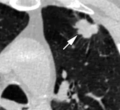
Silicosis on CT

Abnormal CT findings increasingly common among countertop workers

Individuals who work with engineered stone countertops are increasingly being diagnosed with silicosis—a chronic lung condition that is the result of inhaling large amounts of silica dust.

Philips CT 5300

New AI-enabled CT system is officially available in North America

The scanner is equipped with features aimed at addressing some of the biggest pain points of both radiologists and technologists.

Around the web

The new technology shows early potential to make a significant impact on imaging workflows and patient care. 

Richard Heller III, MD, RSNA board member and senior VP of policy at Radiology Partners, offers an overview of policies in Congress that are directly impacting imaging.
 

The two companies aim to improve patient access to high-quality MRI scans by combining their artificial intelligence capabilities.