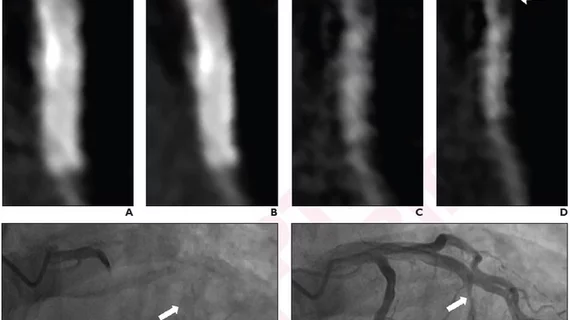
Deep learning, subtraction technique ideal for evaluating stent re-stenosis on coronary CTA
Detecting in-stent restenosis via coronary CTA with hybrid iterative reconstruction (HIR) has historically been an exercise in avoiding false positives, but recently researchers were able to overcome this barrier with a combination of deep learning reconstruction and subtraction images.
The researchers’ efforts were detailed on Aug. 10 in the American Journal of Roentgenology.
In the paper, the experts explained that high rate of false positives produced from standard coronary CTA reconstructions can be blamed on stent-related blooming artifact. To overcome this, they developed a new method of evaluating in-stent restenosis that combined a two-breath-hold (acquisitions both with and without contrast) subtraction technique with deep learning reconstruction (DLR).
From conventional and subtraction images that were reconstructed with both HIR and DLR, two radiologists measured in-stent lumen diameter to assess the presence of restenosis. They did this for 30 patients with an accumulative total of 59 stents.
For the first reader, the combination (conventional and subtraction) with DLR yielded the highest specificity (90.1%), positive predictive value (71.4%), negative predictive value (87%) and accuracy (83.3%).
For the second reader, the same combination with DLR garnered 95.5% specificity, a positive predictive value of 80%, a negative predictive value of 84% and an accuracy of 83.3%. The second reader, however, did achieve similar numbers with the combination images and HIR, and sensitivities were similar with both reconstruction techniques as well.
Corresponding author Yi-Ning Wang, MD, from the Department of Radiology at Peking Union Medical College Hospital in China, and colleagues suggested their findings support the use of the combination with DLR method in the future to identify patients who are suited for more invasive procedures to diagnose and manage restenosis.
“Combination of DLR and subtraction technique yielded optimal diagnostic performance for detecting in-stent restenosis by coronary CTA,” they wrote. “The described technique could guide patient selection for invasive coronary stent evaluation.”
The study abstract can be viewed here.

