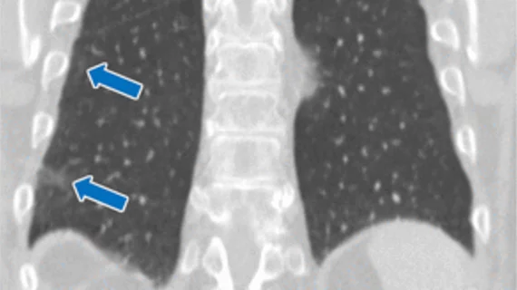
Less than half of incidental interstitial lung abnormalities on CT exams are reported
Nearly half of incidentally detected interstitial lung abnormalities on CT are not reported, according to new data published this week in the journal Radiology.
Interstitial lung abnormalities (ILAs) can signal that a patient is developing interstitial lung disease—a serious condition that causes scarring of the lungs that worsens over time, making it progressively more difficult breathe for many. Outcomes for interstitial lung disease are best when the condition is caught early, as initiating treatment in the earlier stages helps slow disease progression.
This is why any indication of the condition, such as ILAs incidentally identified on imaging, need to be addressed.
“Considering their potential clinical implications, ILAs should be systematically and fully assessed (including evaluation of any disease progression) on every type of CT scan,” co-senior author Mario Silva, from the Department of Medicine and Surgery at University of Parma in Italy, and colleagues cautioned. “Overlooking ILAs (potentially representing treatable fibrotic lung disease) might cause relevant delay in their management.”
However, this is often not the case, as Silve and colleagues' research suggests that these findings are often underreported.
That’s according to the team’s analysis of more than 21,000 patients who underwent either an abdominal or thoracoabdominal CT scans for various indications at a single-center tertiary hospital between January 2008 and December 2015. The group retrospectively examined the scans for the presence of ILAs and compared their own findings to those referenced in the original radiology reports.
Although ILAs were observed in just around 2% of the exams, 44% of the incidental findings, some of which (1%) contained fibrotic features, were not included in the patients’ original reports. According to the patients’ medical records, those with fibrotic ILAs had a four-fold higher risk of respiratory-related mortality. What's more, 39.6% of patients with subpleural ILAs who did not show signs of fibrosis at baseline went on to develop the condition at follow-up.
While the amount of ILAs observed in the study might seem small, the authors described the number of unreported findings as “substantial,” especially considering their consequential nature.
“Despite the discussed attenuating factors, the underreporting rate of the ILAs on these types of body CT scans appeared to be substantial,” the group noted, later adding that these findings “should be systematically reported and fully assessed.”
The study abstract is available here.

