Targeted training reduces certain radiologist errors when reading contrast-enhanced CT
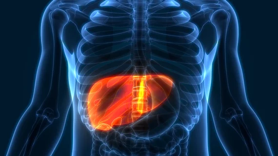
A targeted training approach can help reduce radiologist mistakes when assessing contrast-enhanced CT for liver metastases. However, certain types of errors still persist, researchers detailed in Academic Radiology [1].
Physicians can sometimes overlook low-contrast lesions, such as hepatic metastases or pancreatic adenocarcinoma, on computed tomography scans. Previous research has focused on improved image reconstruction methods or optimized acquisition protocols to fix this blind spot. Scientists with the Mayo Clinic tested a novel education program as a possible solution, producing mixed results.
The change reduced “search errors”—when readers do not gaze at the lesion—but not “classification” mistakes, where radiologists spot the problem but fail to report it as suspicious.
“These results suggest that targeted training can improve performance even for routine tasks, such as low-contrast hepatic metastasis detection, possibly by changing reader interaction patterns with the computer workstation,” Scott S. Hsieh, PhD, with Mayo’s Department of Radiology, and co-authors concluded. “More work is necessary to determine the training methods that will improve classification errors, determine whether reader improvements after targeted training are durable, and compare the effectiveness of targeted training to conventional training.”
The Rochester, Minnesota-based institution recruited 31 radiologists at a single site to take part in the program. They included eight members of the profession who specialize in abdominal imaging, eight more who do not, and 15 senior residents/fellows. Sessions occurred in January and March 2022 and lasted about four hours, including a pretest exam, search and classification training, and another test afterward. For the search aspect, radiologists interpreted up to 30 abdominal CTs, receiving eye-tracker feedback showing where they gazed together with the actual lesion locations. For classification education, meanwhile, radiologists reviewed up to 100 “patches” (20 key CT slices cropped down) and rated lesions on a 100-point scale then weighed them against the findings of expert readers.
Search errors dropped from 11% in the pretest down to 8% after the intervention. However, there was no difference in classification errors nor in the jackknife free-response receiver operator characteristic figure of merit, a measure of diagnostic accuracy that adjusts for reader confidence (difference −0.01, P = .36). Subgroup analysis also revealed that abdominal subspecialists demonstrated no evidence of change.
“It is possible that the dose of classification training delivered here was insufficient to produce a measurable improvement or that classification tasks could be practiced in a way that permits even greater time efficiency and review of more cases (e.g., rating confidence in malignancy for a single image or two sets of images, or delivering training over several shorter sessions to reduce learner fatigue),” the authors noted. “However, our work suggests that search training may be a more promising avenue for improvement than classification training for reducing the rate of missed hepatic metastases.”
Read more in Academic Radiology at the link below.

