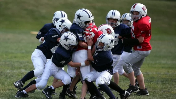
Advanced MRI techniques may provide insight into brain damage stemming from youth sports
Experts have just scanned the first patient involved in new research studying the impact of head injuries on youth athletes.
Conducted at The Podium Institute for Sports Medicine and Technology—part of Oxford University’s Institute of Biomedical Engineering—the study is part of a bigger effort to understand why some children recover better than others following head injuries. CT is typically the imaging modality of choice for children presenting to emergency departments with head injuries, but researchers are instead deploying advanced MRI techniques to study the brain in greater detail.
Experts plan to look at multiple different aspects of brain damage following head injuries, including how it affects nerve fibers, brain metabolism and functional connectivity between different regions of the mind. Imaging findings will be combined with cognitive assessments and patients’ self-reported symptoms to gain objective insight into the trajectory of recovery from both physiological and cognitive standpoints, with the goal of identifying MRI biomarkers that can predict patients’ paths to recovery.
The study will take place over the next 2 and a half years and will include two groups of children—60 who regularly participate in contact sports, including many with either recent or prior head injuries, and 60 controls who do not.
Currently, many unknowns remain related to the long-term impact of head injuries during youth, but experts involved in the study are hopeful that their work can help improve the care and management of such incidents.
“With growing concern regarding a potential link between mild or repetitive traumatic brain injury and long-term cognitive difficulties or even early dementia, there is a pressing need to identify the types of traumatic injuries that may pose a risk,” one of the study’s lead researchers Tim Lawrence, a consultant pediatric neurosurgeon with the Nuffield Department of Clinical Neurosciences at Oxford University, cautioned. “Our study is a step towards better understanding of the mechanisms that underpin damage to the brains of children and adolescents suffering injury.”
“We hope to uncover clinically relevant imaging markers that will turn a difficult-to-see condition into one that can be diagnosed more confidently, and to help clinicians, parents and coaches predict how well a child will recover after a head injury,” Professor Constantin Coussios, Director of the Podium Institute and Director of the Institute of Biomedical Engineering, added later.

