
AI tool detects CVD on cardiac MRI in 20 seconds with high precision
A new artificial intelligence tool can detect heart disease on cardiovascular MRI scans in seconds with equal or superior precision to clinicians.
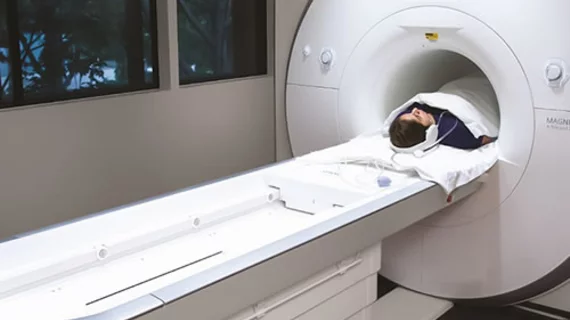
Cardiovascular MRI scans provide vital images that enable providers to measure cardiac structure and function, but interpreting the exams is time consuming and vulnerable to human error. In a recent study published in the Journal of Cardiovascular Magnetic Resonance, experts shared how artificial intelligence could help solve this issue.
“Most existing approaches report measurement accuracy, treating expert analysis (or a consensus thereof) as a truth standard, but this is fundamentally flawed because of the inherent variability and subjectivity of humans,” corresponding author James C. Moon, of the institute of cardiovascular science at University College London, and co-authors explained. “We hypothesized that a carefully trained, fully automated machine learning analysis could be developed and proven to exceed human performance on any CMR scanner and any (non-congenital) disease.”
To test their hypothesis, the researchers trained a machine learning algorithm on 1,923 CMR scans (10 scanner models, 13 institutions, 9 clinical conditions) to segment the left ventricular blood volume and myocardium. The researchers augmented the dataset with an additional 1,277 scans of routine patients to assess the algorithm’s ability to identify mis-segmentations. The algorithm’s performance was then compared to three human readers.
It took the algorithm an average of 20 seconds per patient to analyze the images, which is in stark contrast to the 13 minutes human interpreters required to complete the CMR reads. The quick interpretations did not come at the expense of precision, the researchers noted, as the machine analysis was superior to all three human readers for mis-segmentation.
“We have demonstrated how machine learning can be applied to an important medical problem, cardiac volumetric analysis by CMR, and its performance measured using a clinically important metric—precision,” the experts suggested. “Widespread adoption has the potential to standardize global care, reduce the need for clinical expert time, and significantly reduce sample sizes for clinical trials.”
The experts note that the algorithm is already in use at several institutions in Europe, with plans to implement it more than 40 locations globally within the year.
The detailed research can be viewed in the Journal of Cardiovascular Magnetic Resonance.
More cardiovascular imaging content:
Routine mammograms offer insight into women's risk of cardiovascular disease
CT is a safe, comparable alternative to invasive coronary angiography for chest pain work-ups
Coronary calcium scans benefit firefighters at risk of cardiovascular disease\
Bone density scans show promise for predicting atherosclerotic cardiovascular disease

In addition to her background in journalism, Hannah also has patient-facing experience in clinical settings, having spent more than 12 years working as a registered rad tech. She began covering the medical imaging industry for Innovate Healthcare in 2021.