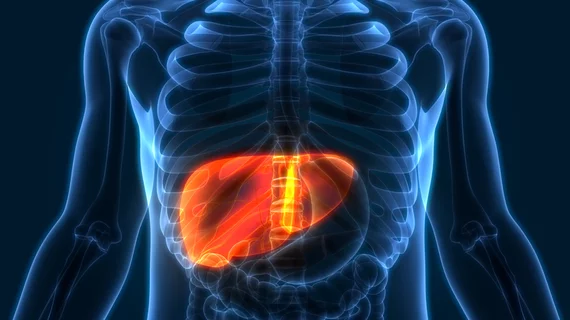
Measurements from functional MRI scans offer non-invasive boost in liver disease assessment
Functional magnetic resonance imaging of the liver is a viable option for assessing chronic liver disease, according to new research.
A new study in the Journal of Hepatology discusses how researchers from MedUni Vienna were able to accurately predict outcomes and complications in patients with chronic liver disease using a risk stratification system that includes functional MRI. By using a combination of functional liver imaging scores (FLIS) derived from gadoxetic acid (GA)-enhanced MRI and splenic diameter measured on MRI, the experts were able to non-invasively assess liver impairment.
FLIS is based on three parameters visualized on GA-MRI: liver enhancement, biliary excretion and portal vein signal intensity. While these scores do offer important predictive assessments, they fall short when it comes to analyzing hepatic decompensation for some patients, including those with both compensated and decompensated advanced chronic liver disease (cACLD and dALCD). The authors noted that spleen size has been previously associated with prognosis in patients with cirrhosis, which is what led them to hypothesize that combining splenic measurements with FLIS might have utility in predicting outcomes in ACLD patients.
“A single examination, GA-MRI, could be used for the diagnostic work-up of liver nodules, as well as for risk stratification for hepatic decompensation and mortality in ACLD,” corresponding author Ahmed Ba-Ssalamah, of Medical University Vienna, General Hospital of Vienna, and co-authors suggested.
This theory was tested in 397 patients with chronic liver disease who had undergone GA-MRI. FLIS was calculated based on imaging, and patients were stratified into three groups based on fibrosis severity and presence/history of hepatic decompensation: Non-ACLD, compensated ACLD (cACLD) and decompensated advanced ACLD (dACLD).
With excellent intra- and inter-reader agreement, splenic diameter was found to be an independent risk factor for hepatic decompensation in cACLD patients. In those same patients, FLIS of 0-3 and splenic diameter of 13 cm were found to have increased risk of decompensation. This was consistent in dACLD patients also, in addition to reliably predicting transplant-free mortality and stratifying the probability of transplant-free survival.
This prompted the authors to suggest that the simple system could be used as a complimentary prognostic tool for assessing liver damage/disease and that it could have a role in future processes once validated.
“The present study confirms and extends the prognostic value of GA-MRI in patients with ACLD by demonstrating the complementary information of FLIS and splenic metrics for stratifying the risks of hepatic decompensation, ACLF, and mortality. Finally, it provides simple, easily applicable algorithms that—after validation—may introduce GA-MRI-based risk stratification into the work-up of patients with ACLD,” the authors concluded.
Related functional MRI articles:
Connectivity abnormalities observed on MRIs of insomnia patients
Combining functional MRI scans with eye tests could help stroke survivors recover lost vision
Even low levels of prenatal alcohol exposure affect brain development, MRI scans show

