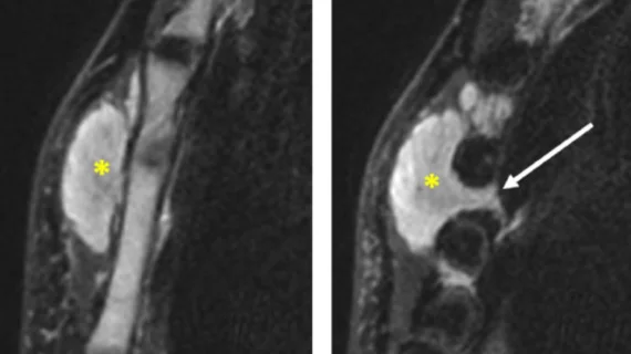
Soft tissue lymphomas versus soft tissue tumors: MRI features reliably differentiate between the two
A new paper published in Academic Radiology highlights experts’ detailed comparison of soft-tissue lymphomas and soft-tissue tumors based on imaging characteristics from MRI scans—an area of study that has not yet been rigorously explored, the authors of the paper indicated.
Corresponding author of the paper, Paolo Spinnato, of the Diagnostic and Interventional Radiology Department with IRCCS Istituto Ortopedico Rizzoli in Italy, and co-authors explained the importance of differentiating between soft-tissue lymphomas (STL) and soft-tissue tumors (STT), suggesting that distinguishing between the two based on imaging could expedite treatment and improve outcomes.
“Faster diagnoses are crucial to rapidly address STL patients to the appropriate referral center, improve outcomes and reduce patients’ anxiety,” the authors wrote. “...the therapeutic strategy would also differ as STLs are treated with systemic therapy alone, whereas curative surgery is recommended for locally advanced STS.”
The experts compared the imaging of 39 patients with STLs to that of 368 patients with other STTs—122 benign STTs and 246 STS. Based on these exams, the researchers were able to narrow down six imaging features that were independent predictors of STLs compared to all other STTs: intermediate SI on T1-WI, homogeneous enhancement (without necrotic areas), no blood signal, no fibrotic signal, no peritumoral enhancement and lack of abnormal intra- and peritumoral vasculature. When these features were present simultaneously, they garnered a sensitivity of .88 and specificity of .88.
The researchers also cited no fat signal to discriminate against STS, infiltrative growth pattern and the vessel and nerve encasement to discriminate against benign STTs as other relevant MRI features.
“These results suggest that conventional MRI can indicate a possible diagnosis of STL. Moreover, clinical and biological features of STL patients appeared poorly specific whereas combining MRI characteristics provided significant diagnostic performances to discriminate from other benign and malignant STTs.”
The experts concluded that radiologists’ awareness of these characteristics could speed up referrals and therapeutic management, improving patient care and outcomes.
The study abstract can be viewed here.

