
Using multiple MRI phases after contrast injection significantly improves visualization of brain tumors
Including multiple delayed MRI sequences after contrast administration could improve visualization of brain tumors.
According to a new paper published in Radiotherapy and Oncology, conducting multiple delayed scans post-contrast injection is beneficial for determining the gross tumor target volume (GTV) for radiation therapy treatments of brain metastases (BM). Experts found that each different time interval offers valuable insight into the size and shape of large volume brain tumors—information that could be used to fine-tune radiotherapy sessions for patients.
“Contrast-enhanced magnetic resonance imaging with gadolinium-based contrast serves as the gold standard for identifying BMs and is the main basis for GTV determination,” corresponding author Yong Yin, with the department of oncology at the Affiliated Hospital of Southwest Medical University in China, and colleagues noted. “However, single-phase static enhancement scans only reflect the instantaneous situation of the contrast agent in the tumor and not the dynamic process of contrast agent penetration and removal, resulting in an incomplete display of BMs boundaries and errors in GTV determination.”
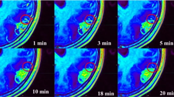
For the study, experts had 155 patients with brain metastases (specifically, lesions 1 centimeter or larger) undergo contrast-enhanced T1-weighted imaging scans at 1-, 3-, 5-, 10-, 18- and 20-minutes post gadolinium-based contrast agent injection. The team compared the volume, shape and signal intensity of the GTVs and brain white matter at each interval to determine if there were optimal delay times.
Starting at the 3-minute delay and continuing through the 20-minute delay, experts recorded an increase in volume of 2.2%, 3.8%, 6.5%, 9.5% and 10.6%, respectively. At the 5-minute interval, the greatest contrast ratio between the GTV and white matter was observed.
“With increasing delay time, contrast agent penetration and removal increase, accelerating longitudinal relaxation and increasing BMs' extent,” the authors explained. “As a result, the early enhancement of tiny metastases is often not obvious compared with that of large-volume BMs, and the degree of enhancement on time-delayed CE scans continues to increase, thus improving the detection rate.”
Previous studies have suggested that scans should begin 10 minutes after contrast injection; however, the differences in the size and shape of the tumors at each individual time interval support inclusion of multiple scan phases, the authors noted. They proposed making additional phases beyond the 10-minute mark mandatory when analyzing the extent of brain metastases.
The study abstract can be viewed here.

In addition to her background in journalism, Hannah also has patient-facing experience in clinical settings, having spent more than 12 years working as a registered rad tech. She began covering the medical imaging industry for Innovate Healthcare in 2021.