MRI shows structural, functional brain abnormalities in Lyme disease long-haulers
Lyme disease long-haulers have structural and functional brain abnormalities, both of which were found to have surprising associations with cognition, according to new research.
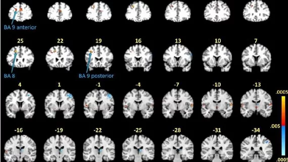
Researchers with Johns Hopkins Medicine utilized functional MR (fMRI) imaging in conjunction with short-term memory tasks to visualize brain activity in patients affected by Lyme disease (12 total), despite having been previously treated for it. Compared to a group of patients (18 total) who had not been previously diagnosed with Lyme disease, those who had been infected displayed unusual activity in the frontal lobe of the brain.
Specifically, the researchers noted activity in the post-treatment Lyme patients’ white matter, which is not normally well visualized on fMRI scans, does not have as much blood flow as gray matter, the study’s lead author lead author Cherie Marvel, PhD, an associate professor of neurology explained in a news release shared by Johns Hopkins Medicine.
“We saw certain areas in the frontal lobe under-activating and others that were over-activating, which was somewhat expected. However, we didn’t see this same white matter activity in the group without post-treatment Lyme,” Marvel shared.
The researchers built upon this finding by completing diffusion tensor imaging (DTI) on the patients. This revealed that the patients with Lyme disease had water leaking along their axons within the same white matter of their frontal lobes.
This finding was surprisingly found to be associated with better cognitive function in patients who had been treated for Lyme. However, these patients also required more time to complete the memory tasks they were given during their scans. The experts suggested that the white matter response could be a sign of adaptive changes but that, ultimately, the structure and function of the brain has been altered by the disease.
The experts acknowledged the small sample size included in their study and suggested that future work should include building on their results by tracking longitudinal changes in white and gray matter. They encouraged the continued use of multimodal imaging.
“Approaching this problem collaboratively with experts in different fields is what gave us these novel findings, and will hopefully answer our remaining questions, specifically about the role of axonal leakage and white matter activation in post-treatment Lyme disease,” Marvel said.
The detailed research is published in PLOS ONE.

