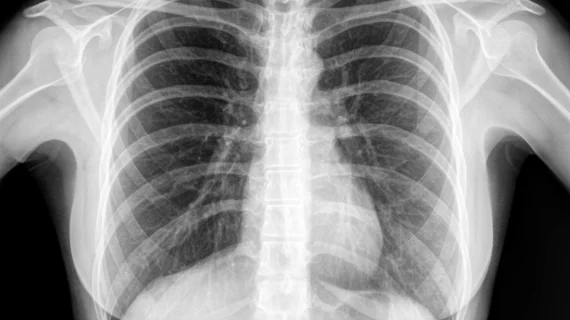
Dynamic chest radiography a suitable, low-cost alternative to V/Q scanning for pulmonary hypertension
The use of dynamic chest radiography (DCR) was recently shown to be comparable to lung ventilation-perfusion (V/Q) scanning for detecting chronic thromboembolic pulmonary hypertension.
Authors of a new paper published in Radiology detailed their recent analysis that did a side-by-side comparison of the two modalities and found that DCR achieved a sensitivity, specificity and accuracy that was in line with that of conventional V/Q scanning when it came to the detection of chronic thromboembolic pulmonary hypertension (CTEPH). The results of the study prompted experts involved in its completion to offer their support to the use of DCR in settings where V/Q scanning might not be readily available.
One of the first steps in diagnosing chronic pulmonary embolism in PH is with a V/Q scan. However, the scan is not widely available outside of academic medical centers, and the use of molybdenum 99 and technetium 99m—radioisotopes that cannot be stored—during isotope shortages threatens the ability to conduct the exam. Furthermore, patients undergoing the exam are subjected to higher doses of ionizing radiation.
For these reasons, corresponding author Yuzo Yamasaki of Kyushu University and colleagues emphasized the need for an option with greater flexibility and less ionizing radiation.
That’s where DCR comes in. DCR is a functional technique that utilizes conventional x-ray technology—a flat-panel detector, pulsed x-ray generator and a special postprocessing station. It captures sequential images during 7 to 10 seconds of breath-holding and does not require contrast media or radionuclides. It also emits a fraction of the radiation dose of typical V/Q scans.
In a cohort of 50 patients with pulmonary hypertension—29 with CTEPH and 21 without—the researchers also found DCR to be an effective diagnostic tool. It achieved sensitivity, specificity and accuracy of 97%, 86%, and 92%. In comparison, these numbers were 100%, 86%, and 94% for V/Q scans.
The authors acknowledged that additional studies are needed to confirm their findings in a clinical setting but suggest that their findings support the use of DCR as an alternative low-cost option in the absence of V/Q scanning technology.
The study abstract is available here.

