New lesion measurement assesses treatment responses more accurately than RECIST
A new way of assessing cancer lesions’ response to treatment could pave the way for developing new cancer therapeutics.
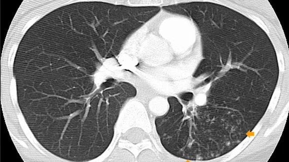
That’s according to a study published recently in Clinical Lung Cancer, where researchers with the University of Colorado Cancer Center on the Anschutz Medical Campus compared a traditional method of measuring lesions on imaging—RECIST—to a newly developed method called MAX [1]. Unlike RECIST, which relies on the largest dimension of a lesion, the MAX method takes two lesion measurements into account and picks the diameter (long or short) that has changed the most since baseline imaging as the greatest indication of treatment efficacy.
Lead author of the study Tami Bang, MD, a thoracic radiologist with the University of Colorado School of Medicine, explained that although RECIST is reliable and time-tested, it is reasonable to consider how other lesion features could change how response rates are assessed.
“We found that we are more consistent with our new MAX method and believe we are more accurately measuring disease,” Bang said in a news release.
This conclusion was derived from the work of Bang and colleagues utilizing the MAX method to assess 249 lung cancer patients with a total of 386 lesions. Researchers also included assessments of 300 additional lung cancer patients with 446 lesions using the RECIST method. All patients in the study underwent CT or PET/CT imaging before and during highly targeted therapies for their cancer.
The experts observed a marked reduction in the impact of lesion location in the chest on apparent effectiveness of targeted therapies. The method was also found to increase the observed response rate overall.
“These results are promising in potentially providing a different way to assess and better develop new treatments for cancer patients,” said senior author of the study Ross Camidge, MD, Joyce Zeff Chair in Lung Cancer Research in the CU Cancer Center, adding that the method could better reflect patients’ responses to therapy.
The authors indicated that future work should focus on the method’s effectiveness when applied to assessments of a wider range of cancers and treatments.
For more information, click here.

