FAPI PET/CT findings linked with risk of sudden cardiac death
Fibroblast activation protein inhibitor (FAPI) uptake on PET/CT imaging was recently found to be associated with increased risks of sudden cardiac death in patients with hypertrophic cardiomyopathy.
The study compared the FAPI PET/CT imaging of patients with hypertrophic cardiomyopathy (HCM) to that of healthy controls and revealed intense and inhomogeneous cardiac FAPI activity in every patient with HCM. Experts involved in the research suggested that these findings indicate a potential role for FAPI PET/CT imaging in detecting changes in myocardial fibrosis, noting that it could be more sensitive to earlier changes than standard cardiac MRI.
“Late gadolinium enhancement reflects changes in extracellular matrix volumes rather than providing targeted imaging of fibroblasts or collagen,” corresponding author Min-Fu Yang, from the Department of Nuclear Medicine at Beijing Chaoyang Hospital in China, and colleagues explained. "Myocardial fibrosis is a highly dynamic process mediated by cardiac fibroblast activation, which manifests as the loss of extracellular matrix homeostasis and excess deposition of collagen.”
FAPI–targeting tracers were developed to detect activated fibroblasts and have been used in PET/CT imaging of cardiovascular diseases, but little is known about the role of cardiac fibroblast activation in HCM. The researchers explored this relationship in 50 patients with HCM in comparison to 22 healthy controls by having both groups undergo cardiac fluorine 18 (18F)–labeled FAPI PET/CT imaging.
Image courtesy of RSNA.
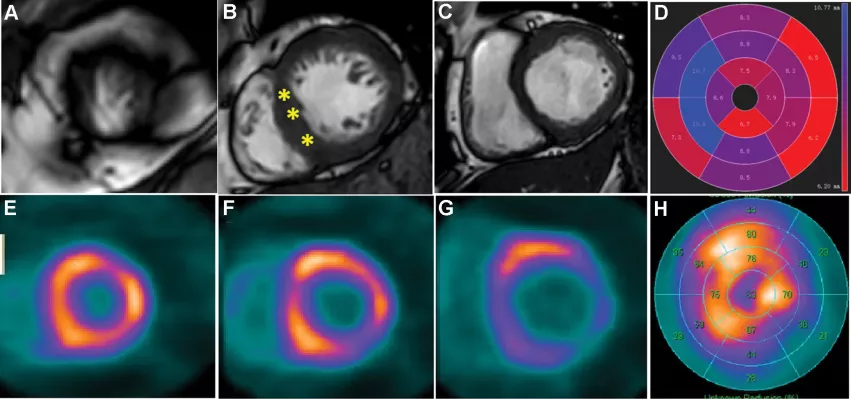
Figure 3: Fluorine 18 (18F)–labeled fibroblast activation protein inhibitor (FAPI) imaging was capable of helping to detect more involved myocardium beyond the hypertrophic region at cardiac MRI in hypertrophic cardiomyopathy (HCM). (A–D) Images in a 52-year-old man with diagnosed HCM for 18 years. The selected short-axis images from cardiac MRI (A–C) showed hypertrophic midseptum (15 mm, *), which is presented with a polar plot (D). (E–H) The corresponding short-axis images of 18F-FAPI (E–G) and polar plot (H) indicated the larger area of cardiac fibroblast activation beyond the hypertrophic region.
Compared to healthy controls, the group with HCM exhibited increased but heterogenous cardiac FAPI activity in the left ventricular myocardium. In the HCM group, 84% of nonhypertrophic segments showed FAPI activity. FAPI amount had a positive relationship left atrial diameter and a negative relationship with left ventricular ejection fraction z-score.
Increased FAPI activity was also shown to be associated with 5-year sudden cardiac death risk scores, a finding that the authors suggested demonstrates the prognostic value of FAPI imaging in risk stratification.
“FAPI imaging may be a promising method in assessing myocardial injury for risk stratification in HCM,” the authors wrote, adding that further research is needed to confirm the role of FAPI imaging in predicting outcomes in patients with HCM.
The study abstract can be viewed in Radiology.


