
How PET imaging could benefit children with cerebral palsy
For the first time, F-18 FDG-PET imaging has been shown to detect brain abnormalities missed on the MRI scans of children with cerebral palsy (CP).
While most children with CP show obvious structural abnormalities on MRI, about 15% do not. But a new study published recently in Frontiers in Neurology found that F-18 FDG-PET scans are capable of identifying patterns of cerebral metabolism changes in patients with otherwise normal imaging [1]. Experts involved in the study suggested that their findings could potentially lead to earlier diagnosis and interventions.
“In the last decade, several studies have explored the structural and functional imaging correlates of impairments in social cognition,” corresponding author of the study Zhijun Pei, PhD, of the Department of Nuclear Medicine and Institute of Anesthesiology and Pain, Taihe Hospital, Hubei University of Medicine in China, and colleagues wrote. "We hypothesized cerebral glucose metabolism in the brain would correlate with GMFCS scores in CP patients. If so, these findings might eventually be helpful in the clinical diagnosis of individual CP patients.”
GMFCS—Gross Motor Function Classification System—scores classify the level of motor function impairment in patients with CP. They are used in conjunction with imaging and other metrics to guide the diagnoses and management of CP.
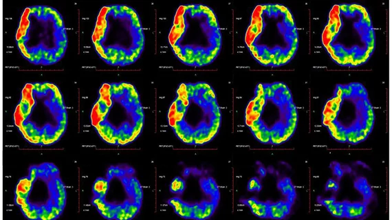
For this study, 31 patients with GMFCS scores of 4 and 5 (indicating worse motor function) who had normal MRI scans underwent F-18 FDG-PET after sedation. Imaging was assessed by two nuclear medicine physicians.
After calculating regional glucose metabolic activity on both the right and left cerebral hemispheres, experts observed asymmetric metabolic reductions in the central region, cerebellum, frontal lobe and parietal lobe.
Researchers then studied whether these values correlated with GMFCS scores. Metabolic activity values of the basal ganglia, left temporal lobe and cerebellum correlated negatively with the scores. When the left cerebellum was put into focus, researchers noted higher metabolic activity values in comparison to the right cerebellum in most patients.
“This marked asymmetric pattern largely reflects physiological developmental changes, rather than structural abnormalities or the clinical presentation of CP,” the authors wrote. “Our results indicate a significant correlation between regional glucose metabolism and the extent of motor impairment, as assessed by GMFCS level.”
The authors added that their findings should encourage further development of their approach in situations when patients with CP do not display structural abnormalities on MRI, as it could help providers differentiate CP associated with different levels of motor impairment.
To view the detailed study, click here.

In addition to her background in journalism, Hannah also has patient-facing experience in clinical settings, having spent more than 12 years working as a registered rad tech. She began covering the medical imaging industry for Innovate Healthcare in 2021.