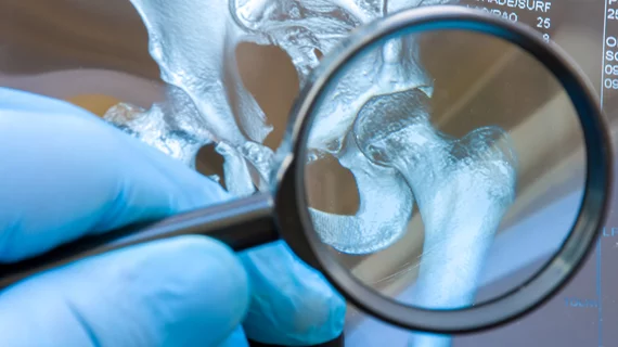
Preoperative radiographic hip measurements predict postoperative complications
Preoperative radiographic measurements of the hip joint could help to inform postoperative outcomes in young orthopaedic patients, according to new research presented today at the American Orthopaedic Society of Sports Medicine 2022 Annual Meeting.
The research centered on patients with femoroacetabular impingement (FAI)—a condition in which extra bone growth occurs in the acetabulum and/or femoral head and causes pain and friction when the bones rub together during movement. Complete resolution of this is achieved via surgery. However, it is not uncommon for patients who undergo surgery for FAI to eventually require an additional operation.
With this in mind, Philip A. Serbin, MD, with Scottish Rite for Children in Dallas, and colleagues hypothesized that preoperative radiographic measurements could hold clues pertaining to postoperative prognosis. For the study, the researchers enrolled 87 patients under the age of 19 who had undergone FAI surgery (56 surgical dislocations vs 31 arthroscopes).
Additional operations were needed in 11.5% of patients after an average of 20.6 months. Serbin and colleagues highlighted three radiographic parameters that increased patients’ risk of reoperation—Lateral Center-Edge Angle (LCEA), Femoro-Epiphyseal Acetabular Roof (FEAR) Index and Sharps angle.
Other key findings:
46% of patients with scores of LCEA <21 required reoperation, compared to 6% with scores above that threshold.
32% of patients with a FEAR index <-8.8 underwent a reoperation compared to 5% of patients >-8.8.
Radiographic signs of hip dysplasia were positively correlated with reoperation.
“Surgeons can utilize these parameters to help in surgical decision making, better predict outcomes and counsel patients about the need for potential subsequent surgery,” Serbin added.
The AOSSM Annual Meeting is taking place in Colorado Springs July 13-17.
Related orthopaedic imaging news:
Do image-guided corticosteroid injections impact COVID risk?
Diagnosing osteoporosis using a deep-radiomics-based approach
Evidence points to optimal MRI sequence for detecting insufficiency fractures
AI tool achieves excellent agreement for knee OA severity classification
Multi-slice knee MRI technique saves time without sacrificing quality
New guidance for knee cartilage MRI seeks to prevent irreversible osteoarthritis

