New guidance for knee cartilage MRI seeks to prevent irreversible osteoarthritis
A leading radiology organization has developed new MRI-based recommendations to help providers better assess and diagnose osteoarthritis.
More than 33% of adults age 65 and older suffer from OA, with related pain, disability and follow-up contributing significant costs to the healthcare system. This new update will help guide more reliable, reproducible results for measuring cartilage degeneration in the knee and, hopefully, prevent irreversible osteoarthritis.
The Radiological Society of North America’s Musculoskeletal Biomarkers Committee of the Quantitative Imaging Biomarkers Alliance (QIBA) published their findings Tuesday in Radiology.
"By the time there is structural damage to the cartilage, treatment choices are very limited," said co-author Majid Chalian, MD, assistant professor of radiology and section head of musculoskeletal imaging and intervention at the University of Washington in Seattle’s Department of Radiology.
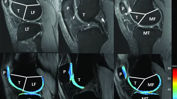
Chalian and co-authors pored over current recommendations and publications for their special report. They noted that two advanced MRI techniques—T1rho and T2 mapping values—are measurable with 3T MRI at 4%-5% variation. A new insight that may help expedite clinical trials, according to the publication.
Additionally, an increase or decrease in T1rho and T2 values of 14% or higher reflects a minimum detectable change, which can be utilized for defining response/progression during quantitative cartilage imaging.
"There was no generally acceptable cut-off for T1rho and T2 values for research and clinical use," Chalian said Sept. 7. "Now we can say that if you image one knee and then do follow-up imaging in two years using the same technique, and you have a 14% change in T1rho and T2 value of the cartilage, that's actually a meaningful change."
It’s a crucial development toward detecting cartilage abnormalities when treatment may still be effective.
The team is hoping they can apply one of the many promising machine learning applications on the market to automatically segment cartilage scans and speed up clinical applications.
RSNA’s QIBA was first launched in 2007 to reduce quantitative imaging variability and advance the use of the technique in clinical trials and everyday practice.

