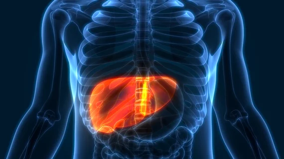
Deep learning decreases CT radiation dose by 65% in patients with liver metastases
Deep learning image reconstruction (DLIR) can significantly reduce the radiation dose for liver cancer patients while also detecting disease spread during abdominal CT scans, according to new research.
The study, published Tuesday in Radiology, assessed the image quality and characterization of liver lesions detected with the reduced dose levels of DLIR compared to the standard doses using filter-back projection (FBJ) contrast-enhanced abdominal CT scans. Historically, using lower dosages to detect liver metastases has deteriorated image quality and lesion detection rates, but new deep learning algorithms have shown promise in compensating for the issue.
“Although reduced-dose imaging is sufficient in many clinical settings, the accuracy of CT in the assessment of small, low-contrast liver lesions quickly becomes inadequate as radiation doses are lowered,” corresponding author Corey T. Jensen, from the Department of Abdominal Imaging at the University of Texas MD Anderson Cancer Center, and co-authors explained.
The prospective study evaluated 161 lesions from 51 patients with biopsy-confirmed colorectal cancer and liver metastases. Each patient underwent two contrast-enhanced abdominal CT scans: one with a reduced dose and one using the standard dose.
The low-dose scans were able to reduce patients’ radiation exposure by 65.1%. As long as the lesions were 0.5 cm or larger, using the lower dose resulted in acceptable image quality. The overall lesion characterization accuracy of DLIR, however, was lower than the standard dose (67.1% versus 80.1%), and had inferior detection capabilities among lesions smaller than 0.5 cm.
DLIR enhanced subjective CT image quality, signal-to-noise ratio and contrast-to-noise ratio compared to the filtered-back projection. But the authors cautioned that more research is still necessary to assess the effectiveness of DLIR for smaller, low-contrast liver lesions.
“Although these improvements from DLIR likely potentiate radiation dose reduction in many clinical scenarios, our results demonstrated that DLIR did not maintain observer lesion detection for very small low-contrast lesions,” the experts warned. “We advise using caution with regard to radiation reduction and the choosing of appropriate radiation dose levels for patients in whom small liver lesion evaluation is important.”
You can view the detailed study in Radiology.

